| 登録情報 | データベース: PDB / ID: 5y53
|
|---|
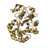
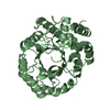
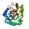
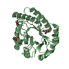
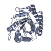
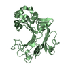
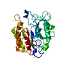
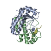
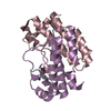
| タイトル | Crystal structure of AL2 PAL domain in complex with AtBMI1b binding site |
|---|
 要素 要素 | - AtBMI1b binding site
- PHD finger protein ALFIN-LIKE 2
|
|---|
 キーワード キーワード | GENE REGULATION / PRC1 interactor / alfin-like family protein / Complex |
|---|
| 機能・相同性 |  機能・相同性情報 機能・相同性情報
response to water deprivation / protein autoubiquitination / transcription coregulator activity / RING-type E3 ubiquitin transferase / ubiquitin-protein transferase activity / chromatin organization / histone binding / transcription cis-regulatory region binding / protein ubiquitination / regulation of DNA-templated transcription ...response to water deprivation / protein autoubiquitination / transcription coregulator activity / RING-type E3 ubiquitin transferase / ubiquitin-protein transferase activity / chromatin organization / histone binding / transcription cis-regulatory region binding / protein ubiquitination / regulation of DNA-templated transcription / zinc ion binding / metal ion binding / nucleus類似検索 - 分子機能 DRIP1/2, RAWUL domain / E3 ubiquitin protein ligase DRIP1-like / Alfin, N-terminal / Alfin1-like, PHD finger / Alfin / Alfin / Zinc finger, C3HC4 type (RING finger) / Zinc finger, PHD-type, conserved site / PHD-finger / Zinc finger, RING-type, conserved site ...DRIP1/2, RAWUL domain / E3 ubiquitin protein ligase DRIP1-like / Alfin, N-terminal / Alfin1-like, PHD finger / Alfin / Alfin / Zinc finger, C3HC4 type (RING finger) / Zinc finger, PHD-type, conserved site / PHD-finger / Zinc finger, RING-type, conserved site / Zinc finger RING-type signature. / Zinc finger PHD-type signature. / Ring finger / Zinc finger PHD-type profile. / Zinc finger, PHD-finger / Zinc finger, PHD-type / PHD zinc finger / Zinc finger, FYVE/PHD-type / Zinc finger RING-type profile. / Zinc finger, RING-type / Zinc finger, RING/FYVE/PHD-type類似検索 - ドメイン・相同性 E3 ubiquitin protein ligase DRIP1 / PHD finger protein ALFIN-LIKE 2類似検索 - 構成要素 |
|---|
| 生物種 |   Arabidopsis thaliana (シロイヌナズナ) Arabidopsis thaliana (シロイヌナズナ) |
|---|
| 手法 |  X線回折 / シンクロトロン / 分子置換 / 解像度: 1.598 Å X線回折 / シンクロトロン / 分子置換 / 解像度: 1.598 Å |
|---|
 データ登録者 データ登録者 | Peng, L. / Wang, L.L. / Huang, Y. |
|---|
 引用 引用 | ジャーナル: J. Mol. Biol. / 年: 2018
タイトル: Structural Analysis of the Arabidopsis AL2-PAL and PRC1 Complex Provides Mechanistic Insight into Active-to-Repressive Chromatin State Switch
著者: Peng, L. / Wang, L. / Zhang, Y. / Dong, A. / Shen, W.H. / Huang, Y. |
|---|
| 履歴 | | 登録 | 2017年8月7日 | 登録サイト: PDBJ / 処理サイト: PDBJ |
|---|
| 改定 1.0 | 2018年8月15日 | Provider: repository / タイプ: Initial release |
|---|
| 改定 1.1 | 2018年9月19日 | Group: Data collection / Structure summary / カテゴリ: entity / entity_name_com / struct
Item: _entity.pdbx_description / _entity_name_com.name ..._entity.pdbx_description / _entity_name_com.name / _struct.pdbx_descriptor / _struct.title |
|---|
| 改定 1.2 | 2019年1月16日 | Group: Data collection / Database references / カテゴリ: citation / citation_author
Item: _citation.country / _citation.journal_abbrev ..._citation.country / _citation.journal_abbrev / _citation.journal_id_ASTM / _citation.journal_id_CSD / _citation.journal_id_ISSN / _citation.journal_volume / _citation.page_first / _citation.page_last / _citation.pdbx_database_id_DOI / _citation.pdbx_database_id_PubMed / _citation.title / _citation.year |
|---|
| 改定 1.3 | 2023年11月22日 | Group: Data collection / Database references / Refinement description
カテゴリ: chem_comp_atom / chem_comp_bond ...chem_comp_atom / chem_comp_bond / database_2 / pdbx_initial_refinement_model
Item: _database_2.pdbx_DOI / _database_2.pdbx_database_accession |
|---|
|
|---|
 ムービー
ムービー コントローラー


 データを開く
 基本情報
基本情報 構造の表示
構造の表示 ダウンロードとリンク
 その他のダウンロード
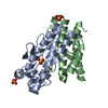


 PDBj
PDBj

 集合体
集合体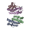

 分子量: 18.015 Da / 分子数: 753 / 由来タイプ: 天然 / 式: H2O
 試料調製
試料調製 / ビームライン: BL19U1 / 波長: 0.97853 Å
 解析
解析