 Movie
Movie Controller

[English] 日本語
 Yorodumi
- PDB-5y21: Crystal structure of AL2 PAL domain in complex with AtRing1a prox... -
+ Open data

- Basic information
| Entry | Database: PDB / ID: 5y21 | ||||||
|---|---|---|---|---|---|---|---|
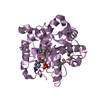
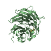
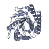
| Title | Crystal structure of AL2 PAL domain in complex with AtRing1a proximal site | ||||||
 Components Components |
| ||||||
 Keywords Keywords | GENE REGULATION / PRC1 interactor / Alfin-like family protein / Complex | ||||||
| Function / homology |  Function and homology information Function and homology informationmaintenance of floral meristem identity / maintenance of inflorescence meristem identity / maintenance of shoot apical meristem identity / seed germination / PRC1 complex / root development / cell fate determination / negative regulation of gene expression, epigenetic / transcription coregulator activity / RING-type E3 ubiquitin transferase ...maintenance of floral meristem identity / maintenance of inflorescence meristem identity / maintenance of shoot apical meristem identity / seed germination / PRC1 complex / root development / cell fate determination / negative regulation of gene expression, epigenetic / transcription coregulator activity / RING-type E3 ubiquitin transferase / ubiquitin protein ligase activity / chromatin organization / histone binding / transcription cis-regulatory region binding / protein ubiquitination / negative regulation of DNA-templated transcription / regulation of DNA-templated transcription / zinc ion binding / nucleus Similarity search - Function | ||||||
| Biological species |  | ||||||
| Method |  X-RAY DIFFRACTION / SYNCHROTRON / MOLECULAR REPLACEMENT / Resolution: 1.769 Å X-RAY DIFFRACTION / SYNCHROTRON / MOLECULAR REPLACEMENT / Resolution: 1.769 Å | ||||||
 Authors Authors | Peng, L. / Wang, L.L. / Huang, Y. | ||||||
 Citation Citation | Journal: J. Mol. Biol. / Year: 2018 Title: Structural Analysis of the Arabidopsis AL2-PAL and PRC1 Complex Provides Mechanistic Insight into Active-to-Repressive Chromatin State Switch Authors: Peng, L. / Wang, L. / Zhang, Y. / Dong, A. / Shen, W.H. / Huang, Y. | ||||||
| History |
|
- Structure visualization
| Structure viewer | Molecule: MolmilJmol/JSmol |
|---|
- Downloads & links
-Download
| PDBx/mmCIF format | 5y21.cif.gz | 137.6 KB | Display | PDBx/mmCIF format |
|---|---|---|---|---|
| PDB format | pdb5y21.ent.gz | 105.9 KB | Display | PDB format |
| PDBx/mmJSON format | 5y21.json.gz | Tree view | PDBx/mmJSON format | |
| Others |  Other downloads Other downloads |
-Validation report
| Arichive directory | https://data.pdbj.org/pub/pdb/validation_reports/y2/5y21ftp://data.pdbj.org/pub/pdb/validation_reports/y2/5y21 | HTTPS FTP |
|---|
-Related structure data
| Related structure data | 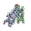 5xvjC 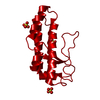 5xvlSC 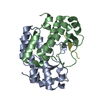 5xvwC 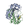 5y53C S: Starting model for refinement C: citing same article ( |
|---|---|
| Similar structure data |


-Links
 PDBj
PDBj

- Assembly
| Deposited unit | 
| ||||||||
|---|---|---|---|---|---|---|---|---|---|
| 1 |
| ||||||||
| Unit cell |
|
-Components
| #1: Protein | Mass: 15508.561 Da / Num. of mol.: 2 / Fragment: UNP residues 10-142 Source method: isolated from a genetically manipulated source Source: (gene. exp.)  #2: Protein/peptide | Mass: 1823.156 Da / Num. of mol.: 2 / Fragment: UNP residues 307-320 / Source method: obtained synthetically / Source: (synth.) #3: Water | ChemComp-HOH / |  Mass: 18.015 Da / Num. of mol.: 487 / Source method: isolated from a natural source / Formula: H2O Mass: 18.015 Da / Num. of mol.: 487 / Source method: isolated from a natural source / Formula: H2O |
|---|
-Experimental details
-Experiment
| Experiment | Method: X-RAY DIFFRACTION / Number of used crystals: 1 |
|---|
- Sample preparation
| Crystal | Density Matthews: 2.39 Å3/Da / Density % sol: 48.62 % |
|---|---|
| Crystal grow | Temperature: 289 K / Method: vapor diffusion, hanging drop / pH: 7.5 / Details: 25% PEG2000 MME, 0.1 M Tris, pH 7.5 |
-Data collection
| Diffraction | Mean temperature: 100 K |
|---|---|
| Diffraction source | Source: SYNCHROTRON / Site: SSRF  / Beamline: BL19U1 / Wavelength: 0.97907 Å / Beamline: BL19U1 / Wavelength: 0.97907 Å |
| Detector | Type: DECTRIS PILATUS 6M / Detector: PIXEL / Date: Jun 27, 2013 |
| Radiation | Protocol: SINGLE WAVELENGTH / Monochromatic (M) / Laue (L): M / Scattering type: x-ray |
| Radiation wavelength | Wavelength: 0.97907 Å / Relative weight: 1 |
| Reflection | Resolution: 1.769→50 Å / Num. obs: 31734 / % possible obs: 99.3 % / Redundancy: 3.7 % / Net I/σ(I): 35.4 |
| Reflection shell | Resolution: 1.77→1.83 Å / Redundancy: 3.7 % / Rmerge(I) obs: 0.154 / Mean I/σ(I) obs: 14.4 / Num. unique obs: 3179 / % possible all: 100 |
- Processing
| Software |
| |||||||||||||||||||||||||||||||||||||||||||||||||||||||||||||||||||||||||||||||||||||||||||||||||||||||||
|---|---|---|---|---|---|---|---|---|---|---|---|---|---|---|---|---|---|---|---|---|---|---|---|---|---|---|---|---|---|---|---|---|---|---|---|---|---|---|---|---|---|---|---|---|---|---|---|---|---|---|---|---|---|---|---|---|---|---|---|---|---|---|---|---|---|---|---|---|---|---|---|---|---|---|---|---|---|---|---|---|---|---|---|---|---|---|---|---|---|---|---|---|---|---|---|---|---|---|---|---|---|---|---|---|---|---|
| Refinement | Method to determine structure: MOLECULAR REPLACEMENT Starting model: 5XVL Resolution: 1.769→27.791 Å / SU ML: 0.15 / Cross valid method: FREE R-VALUE / σ(F): 1.4 / Phase error: 17.96
| |||||||||||||||||||||||||||||||||||||||||||||||||||||||||||||||||||||||||||||||||||||||||||||||||||||||||
| Solvent computation | Shrinkage radii: 0.9 Å / VDW probe radii: 1.11 Å | |||||||||||||||||||||||||||||||||||||||||||||||||||||||||||||||||||||||||||||||||||||||||||||||||||||||||
| Refinement step | Cycle: LAST / Resolution: 1.769→27.791 Å
| |||||||||||||||||||||||||||||||||||||||||||||||||||||||||||||||||||||||||||||||||||||||||||||||||||||||||
| Refine LS restraints |
| |||||||||||||||||||||||||||||||||||||||||||||||||||||||||||||||||||||||||||||||||||||||||||||||||||||||||
| LS refinement shell |
|
