 Movie
Movie Controller

[English] 日本語
 Yorodumi
- PDB-4f2u: Structure of the N254Y/H258Y double mutant of the Phosphatidylino... -
+ Open data

- Basic information
| Entry | Database: PDB / ID: 4f2u | ||||||
|---|---|---|---|---|---|---|---|
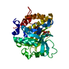
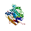
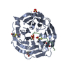
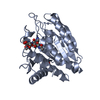
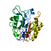
| Title | Structure of the N254Y/H258Y double mutant of the Phosphatidylinositol-Specific Phospholipase C from S.aureus | ||||||
 Components Components | 1-phosphatidylinositol phosphodiesterase | ||||||
 Keywords Keywords | LYASE / alpha beta barrel / Phosphatidylinositol-specific phospholipase C / membrane interface | ||||||
| Function / homology |  Function and homology information Function and homology informationphosphatidylinositol diacylglycerol-lyase / phosphatidylinositol diacylglycerol-lyase activity / phosphoric diester hydrolase activity / lipid catabolic process / extracellular region Similarity search - Function | ||||||
| Biological species |   Staphylococcus aureus subsp. aureus (bacteria) Staphylococcus aureus subsp. aureus (bacteria) | ||||||
| Method |  X-RAY DIFFRACTION / MOLECULAR REPLACEMENT / Resolution: 2.19 Å X-RAY DIFFRACTION / MOLECULAR REPLACEMENT / Resolution: 2.19 Å | ||||||
 Authors Authors | Cheng, J. / Goldstein, R. / Stec, B. / Gershenson, A. / Roberts, M.F. | ||||||
 Citation Citation | Journal: J.Biol.Chem. / Year: 2012 Title: Competition between Anion Binding and Dimerization Modulates Staphylococcus aureus Phosphatidylinositol-specific Phospholipase C Enzymatic Activity. Authors: Cheng, J. / Goldstein, R. / Stec, B. / Gershenson, A. / Roberts, M.F. | ||||||
| History |
|
- Structure visualization
| Structure viewer | Molecule: MolmilJmol/JSmol |
|---|
- Downloads & links
-Download
| PDBx/mmCIF format | 4f2u.cif.gz | 81.3 KB | Display | PDBx/mmCIF format |
|---|---|---|---|---|
| PDB format | pdb4f2u.ent.gz | 59.4 KB | Display | PDB format |
| PDBx/mmJSON format | 4f2u.json.gz | Tree view | PDBx/mmJSON format | |
| Others |  Other downloads Other downloads |
-Validation report
| Arichive directory | https://data.pdbj.org/pub/pdb/validation_reports/f2/4f2uftp://data.pdbj.org/pub/pdb/validation_reports/f2/4f2u | HTTPS FTP |
|---|
-Related structure data
| Related structure data | 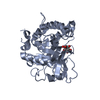 4f2bC 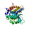 4f2tC 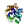 3v18S S: Starting model for refinement C: citing same article ( |
|---|---|
| Similar structure data |


-Links
 PDBj
PDBj- Assembly
| Deposited unit | 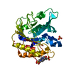
| ||||||||
|---|---|---|---|---|---|---|---|---|---|
| 1 |
| ||||||||
| Unit cell |
|
-Components
| #1: Protein | Mass: 35316.238 Da / Num. of mol.: 1 / Fragment: UNP residues 11-312 / Mutation: N254Y/H258Y Source method: isolated from a genetically manipulated source Source: (gene. exp.) Staphylococcus aureus subsp. aureus (bacteria)Strain: Newman / Gene: plc, NWMN_0041 / Production host: References: UniProt: P45723, phosphatidylinositol diacylglycerol-lyase | ||||
|---|---|---|---|---|---|
| #2: Chemical |   Mass: 238.305 Da / Num. of mol.: 2 / Source method: obtained synthetically / Formula: C8H18N2O4S / Comment: pH buffer*YM Mass: 238.305 Da / Num. of mol.: 2 / Source method: obtained synthetically / Formula: C8H18N2O4S / Comment: pH buffer*YM#3: Chemical | ChemComp-SO4 / |   Mass: 96.063 Da / Num. of mol.: 1 / Source method: obtained synthetically / Formula: SO4 Mass: 96.063 Da / Num. of mol.: 1 / Source method: obtained synthetically / Formula: SO4#4: Water | ChemComp-HOH / |  Mass: 18.015 Da / Num. of mol.: 288 / Source method: isolated from a natural source / Formula: H2O Mass: 18.015 Da / Num. of mol.: 288 / Source method: isolated from a natural source / Formula: H2O |
-Experimental details
-Experiment
| Experiment | Method: X-RAY DIFFRACTION / Number of used crystals: 1 |
|---|
- Sample preparation
| Crystal | Density Matthews: 2.11 Å3/Da / Density % sol: 41.81 % |
|---|---|
| Crystal grow | Temperature: 293 K / Method: vapor diffusion, hanging drop / pH: 7.5 Details: 22% PEG 4000,0.100 M Hepes, 10% isopropanol, pH 7.5, VAPOR DIFFUSION, HANGING DROP, temperature 293K |
-Data collection
| Diffraction | Mean temperature: 120 K |
|---|---|
| Diffraction source | Source: ROTATING ANODE / Type: RIGAKU MICROMAX-007 HF / Wavelength: 1.54 Å |
| Detector | Type: RIGAKU RAXIS IV++ / Detector: IMAGE PLATE / Date: Aug 15, 2011 / Details: Osmic VariMax |
| Radiation | Monochromator: Osmic VariMax / Protocol: SINGLE WAVELENGTH / Monochromatic (M) / Laue (L): M / Scattering type: x-ray |
| Radiation wavelength | Wavelength: 1.54 Å / Relative weight: 1 |
| Reflection | Resolution: 2.19→50 Å / Num. all: 94114 / % possible obs: 99.9 % / Observed criterion σ(F): 0 / Observed criterion σ(I): -3 / Redundancy: 6 % / Rmerge(I) obs: 0.117 / Rsym value: 0.117 / Net I/σ(I): 26.49 |
| Reflection shell | Resolution: 2.19→2.24 Å / Redundancy: 6 % / % possible all: 100 |
- Processing
| Software |
| |||||||||||||||||||||||||||||||||||||||||||||||||
|---|---|---|---|---|---|---|---|---|---|---|---|---|---|---|---|---|---|---|---|---|---|---|---|---|---|---|---|---|---|---|---|---|---|---|---|---|---|---|---|---|---|---|---|---|---|---|---|---|---|---|
| Refinement | Method to determine structure: MOLECULAR REPLACEMENT Starting model: PDB entry 3V18 Resolution: 2.19→30.854 Å / SU ML: 0.3 / σ(F): 1.34 / Phase error: 20.98 / Stereochemistry target values: ML
| |||||||||||||||||||||||||||||||||||||||||||||||||
| Solvent computation | Shrinkage radii: 0.98 Å / VDW probe radii: 1.2 Å / Solvent model: FLAT BULK SOLVENT MODEL / Bsol: 32.447 Å2 / ksol: 0.3 e/Å3 | |||||||||||||||||||||||||||||||||||||||||||||||||
| Displacement parameters |
| |||||||||||||||||||||||||||||||||||||||||||||||||
| Refinement step | Cycle: LAST / Resolution: 2.19→30.854 Å
| |||||||||||||||||||||||||||||||||||||||||||||||||
| Refine LS restraints |
| |||||||||||||||||||||||||||||||||||||||||||||||||
| LS refinement shell |
|
