 Movie
Movie Controller

[English] 日本語
 Yorodumi
- PDB-4gga: Structural Analysis of Human Cdc20 Supports Multi-site Degron Rec... -
+ Open data

- Basic information
| Entry | Database: PDB / ID: 4gga | ||||||
|---|---|---|---|---|---|---|---|
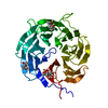
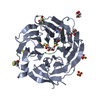
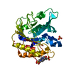
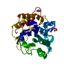
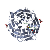
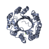
| Title | Structural Analysis of Human Cdc20 Supports Multi-site Degron Recognition by APC/C | ||||||
 Components Components | Cell division cycle protein 20 homolog | ||||||
 Keywords Keywords | CELL CYCLE / mitosis / securin / ubiquitination / WD40 | ||||||
| Function / homology |  Function and homology information Function and homology informationmetaphase/anaphase transition of cell cycle / metaphase/anaphase transition of meiosis I / Inhibition of the proteolytic activity of APC/C required for the onset of anaphase by mitotic spindle checkpoint components / mitotic checkpoint complex / positive regulation of ubiquitin protein ligase activity / positive regulation of anaphase-promoting complex-dependent catabolic process / positive regulation of synapse maturation / regulation of meiotic nuclear division / Conversion from APC/C:Cdc20 to APC/C:Cdh1 in late anaphase / Inactivation of APC/C via direct inhibition of the APC/C complex ...metaphase/anaphase transition of cell cycle / metaphase/anaphase transition of meiosis I / Inhibition of the proteolytic activity of APC/C required for the onset of anaphase by mitotic spindle checkpoint components / mitotic checkpoint complex / positive regulation of ubiquitin protein ligase activity / positive regulation of anaphase-promoting complex-dependent catabolic process / positive regulation of synapse maturation / regulation of meiotic nuclear division / Conversion from APC/C:Cdc20 to APC/C:Cdh1 in late anaphase / Inactivation of APC/C via direct inhibition of the APC/C complex / APC/C:Cdc20 mediated degradation of mitotic proteins / regulation of dendrite development / anaphase-promoting complex / Phosphorylation of Emi1 / anaphase-promoting complex-dependent catabolic process / regulation of meiotic cell cycle / anaphase-promoting complex binding / positive regulation of synaptic plasticity / positive regulation of mitotic metaphase/anaphase transition / ubiquitin ligase activator activity / mitotic sister chromatid cohesion / mitotic spindle assembly checkpoint signaling / Regulation of APC/C activators between G1/S and early anaphase / mitotic spindle assembly / ubiquitin-like ligase-substrate adaptor activity / Amplification of signal from unattached kinetochores via a MAD2 inhibitory signal / regulation of mitotic cell cycle / Mitotic Prometaphase / EML4 and NUDC in mitotic spindle formation / APC/C:Cdc20 mediated degradation of Cyclin B / APC-Cdc20 mediated degradation of Nek2A / Resolution of Sister Chromatid Cohesion / APC/C:Cdc20 mediated degradation of Securin / SCF-beta-TrCP mediated degradation of Emi1 / Cdc20:Phospho-APC/C mediated degradation of Cyclin A / RHO GTPases Activate Formins / APC/C:Cdh1 mediated degradation of Cdc20 and other APC/C:Cdh1 targeted proteins in late mitosis/early G1 / kinetochore / spindle / histone deacetylase binding / spindle pole / Separation of Sister Chromatids / nervous system development / Antigen processing: Ubiquitination & Proteasome degradation / cell differentiation / Ub-specific processing proteases / protein ubiquitination / cell division / positive regulation of cell population proliferation / centrosome / perinuclear region of cytoplasm / nucleoplasm / cytosol Similarity search - Function | ||||||
| Biological species |  Homo sapiens (human) Homo sapiens (human) | ||||||
| Method |  X-RAY DIFFRACTION / SYNCHROTRON / MOLECULAR REPLACEMENT / molecular replacement / Resolution: 2.044 Å X-RAY DIFFRACTION / SYNCHROTRON / MOLECULAR REPLACEMENT / molecular replacement / Resolution: 2.044 Å | ||||||
 Authors Authors | Luo, X. / Tian, W. / Tomchick, D.R. | ||||||
 Citation Citation | Journal: Proc.Natl.Acad.Sci.USA / Year: 2012 Title: Structural analysis of human Cdc20 supports multisite degron recognition by APC/C. Authors: Tian, W. / Li, B. / Warrington, R. / Tomchick, D.R. / Yu, H. / Luo, X. #1: Journal: Mol.Cell / Year: 2007Title: Cdc20: a WD40 activator for a cell cycle degradation machine Authors: Yu, H. #2: Journal: J.Biol.Chem. / Year: 2007Title: KEN-box-dependent degradation of Bub1 spindle checkpoint kinase by the anaphase-promoting complex/cyclosome Authors: Qi, W. / Yu, H. #3: Journal: Dev.Cell / Year: 2001Title: Mad2-independent inhibition of APC-Cdc20 by the mitotic checkpoint protein BubR1 Authors: Tang, Z. / Bharadwaj, R. / Li, B. / Yu, H. | ||||||
| History |
|
- Structure visualization
| Structure viewer | Molecule: MolmilJmol/JSmol |
|---|
- Downloads & links
-Download
| PDBx/mmCIF format | 4gga.cif.gz | 188.8 KB | Display | PDBx/mmCIF format |
|---|---|---|---|---|
| PDB format | pdb4gga.ent.gz | 151.5 KB | Display | PDB format |
| PDBx/mmJSON format | 4gga.json.gz | Tree view | PDBx/mmJSON format | |
| Others |  Other downloads Other downloads |
-Validation report
| Arichive directory | https://data.pdbj.org/pub/pdb/validation_reports/gg/4ggaftp://data.pdbj.org/pub/pdb/validation_reports/gg/4gga | HTTPS FTP |
|---|
-Related structure data


-Links
 PDBj
PDBj


- Assembly
| Deposited unit | 
| ||||||||
|---|---|---|---|---|---|---|---|---|---|
| 1 |
| ||||||||
| Unit cell |
|
-Components
| #1: Protein | Mass: 46350.996 Da / Num. of mol.: 1 Source method: isolated from a genetically manipulated source Source: (gene. exp.) Homo sapiens (human) / Gene: CDC20 / Plasmid: pFastBac / Production host:   Spodoptera frugiperda (fall armyworm) / References: UniProt: Q12834 Spodoptera frugiperda (fall armyworm) / References: UniProt: Q12834 |
|---|---|
| #2: Water | ChemComp-HOH /  Mass: 18.015 Da / Num. of mol.: 247 / Source method: isolated from a natural source / Formula: H2O Mass: 18.015 Da / Num. of mol.: 247 / Source method: isolated from a natural source / Formula: H2O |
-Experimental details
-Experiment
| Experiment | Method: X-RAY DIFFRACTION / Number of used crystals: 1 |
|---|
- Sample preparation
| Crystal | Density Matthews: 2.13 Å3/Da / Density % sol: 42.36 % |
|---|---|
| Crystal grow | Temperature: 293 K / Method: vapor diffusion, sitting drop / pH: 5.6 Details: 100 mM Na citrate, 15 % (w/v) PEG 6000, and 4% MPD, pH 5.6, VAPOR DIFFUSION, SITTING DROP, temperature 293K |
-Data collection
| Diffraction | Mean temperature: 100 K |
|---|---|
| Diffraction source | Source: SYNCHROTRON / Site: APS  / Beamline: 19-ID / Wavelength: 0.97937 Å / Beamline: 19-ID / Wavelength: 0.97937 Å |
| Detector | Type: ADSC QUANTUM 315r / Detector: CCD / Date: Apr 18, 2010 / Details: monochromator |
| Radiation | Monochromator: SAGITALLY FOCUSED Si(111) / Protocol: SINGLE WAVELENGTH / Monochromatic (M) / Laue (L): M / Scattering type: x-ray |
| Radiation wavelength | Wavelength: 0.97937 Å / Relative weight: 1 |
| Reflection | Resolution: 2.044→68.589 Å / Num. all: 25867 / Num. obs: 24684 / % possible obs: 95.4 % / Observed criterion σ(F): 0 / Observed criterion σ(I): 0 / Redundancy: 5.6 % / Biso Wilson estimate: 18.7 Å2 / Limit h max: 19 / Limit h min: 0 / Limit k max: 42 / Limit k min: 0 / Limit l max: 54 / Limit l min: 0 / Rmerge(I) obs: 0.222 / Net I/σ(I): 10 |
| Reflection scale | Group code: 1 |
| Reflection shell | Resolution: 2.05→2.09 Å / Redundancy: 2.7 % / Rmerge(I) obs: 0.599 / Num. unique all: 1044 / % possible all: 83.8 |
-Phasing
| Phasing | Method: molecular replacement |
|---|
- Processing
| Software |
| ||||||||||||||||||||||||||||||||||||||||||||||||||||||||||||||||||||||||||||||||||||||||||||||||||||
|---|---|---|---|---|---|---|---|---|---|---|---|---|---|---|---|---|---|---|---|---|---|---|---|---|---|---|---|---|---|---|---|---|---|---|---|---|---|---|---|---|---|---|---|---|---|---|---|---|---|---|---|---|---|---|---|---|---|---|---|---|---|---|---|---|---|---|---|---|---|---|---|---|---|---|---|---|---|---|---|---|---|---|---|---|---|---|---|---|---|---|---|---|---|---|---|---|---|---|---|---|---|
| Refinement | Method to determine structure: MOLECULAR REPLACEMENT / Resolution: 2.044→38.347 Å / Occupancy max: 1 / Occupancy min: 0.5 / SU ML: 0.2 / Cross valid method: THROUGHOUT / σ(F): 0 / σ(I): 0 / Phase error: 19.41 / Stereochemistry target values: Engh & Huber
| ||||||||||||||||||||||||||||||||||||||||||||||||||||||||||||||||||||||||||||||||||||||||||||||||||||
| Solvent computation | Shrinkage radii: 0.9 Å / VDW probe radii: 1.11 Å / Solvent model: FLAT BULK SOLVENT MODEL | ||||||||||||||||||||||||||||||||||||||||||||||||||||||||||||||||||||||||||||||||||||||||||||||||||||
| Displacement parameters | Biso max: 139.81 Å2 / Biso mean: 23.3523 Å2 / Biso min: 3.56 Å2 | ||||||||||||||||||||||||||||||||||||||||||||||||||||||||||||||||||||||||||||||||||||||||||||||||||||
| Refine analyze | Luzzati sigma a obs: 0.2 Å | ||||||||||||||||||||||||||||||||||||||||||||||||||||||||||||||||||||||||||||||||||||||||||||||||||||
| Refinement step | Cycle: LAST / Resolution: 2.044→38.347 Å
| ||||||||||||||||||||||||||||||||||||||||||||||||||||||||||||||||||||||||||||||||||||||||||||||||||||
| Refine LS restraints |
| ||||||||||||||||||||||||||||||||||||||||||||||||||||||||||||||||||||||||||||||||||||||||||||||||||||
| LS refinement shell | Refine-ID: X-RAY DIFFRACTION / Total num. of bins used: 9
| ||||||||||||||||||||||||||||||||||||||||||||||||||||||||||||||||||||||||||||||||||||||||||||||||||||
| Refinement TLS params. | Method: refined / Refine-ID: X-RAY DIFFRACTION
| ||||||||||||||||||||||||||||||||||||||||||||||||||||||||||||||||||||||||||||||||||||||||||||||||||||
| Refinement TLS group |
|
