 Movie
Movie Controller

[English] 日本語
 Yorodumi
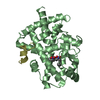
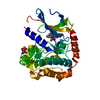
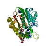
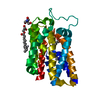
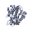
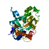
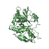
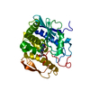
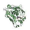
- PDB-1l1e: Crystal Structure of Mycolic Acid Cyclopropane Synthase PcaA Comp... -
+ Open data

- Basic information
| Entry | Database: PDB / ID: 1l1e | ||||||
|---|---|---|---|---|---|---|---|
| Title | Crystal Structure of Mycolic Acid Cyclopropane Synthase PcaA Complexed with S-adenosyl-L-homocysteine | ||||||
 Components Components | mycolic acid synthase | ||||||
 Keywords Keywords | TRANSFERASE / METHYLTRANSFERASE / S-ADENOSYL-L-HOMOCYSTEINE COFACTOR / ALPHA/BETA / Structural Genomics / PSI / Protein Structure Initiative / TB Structural Genomics Consortium / TBSGC | ||||||
| Function / homology |  Function and homology information Function and homology informationcyclopropane-fatty-acyl-phospholipid synthase / cyclopropane-fatty-acyl-phospholipid synthase activity / symbiont-mediated perturbation of host innate immune response / S-adenosylmethionine metabolic process / mycolic acid biosynthetic process / symbiont-mediated evasion of host immune response / lipid biosynthetic process / methylation / cytoplasm Similarity search - Function | ||||||
| Biological species |   Mycobacterium tuberculosis (bacteria) Mycobacterium tuberculosis (bacteria) | ||||||
| Method |  X-RAY DIFFRACTION / SYNCHROTRON / MAD / Resolution: 2 Å X-RAY DIFFRACTION / SYNCHROTRON / MAD / Resolution: 2 Å | ||||||
 Authors Authors | Huang, C.-C. / Smith, C.V. / Glickman, M.S. / Jacobs Jr., W.R. / Sacchettini, J.C. / TB Structural Genomics Consortium (TBSGC) | ||||||
 Citation Citation | Journal: J.Biol.Chem. / Year: 2002 Title: Crystal structures of mycolic acid cyclopropane synthases from Mycobacterium tuberculosis. Authors: Huang, C.-C. / Smith, C.V. / Glickman, M.S. / Jacobs Jr., W.R. / Sacchettini, J.C. | ||||||
| History |
|
- Structure visualization
| Structure viewer | Molecule: MolmilJmol/JSmol |
|---|
- Downloads & links
-Download
| PDBx/mmCIF format | 1l1e.cif.gz | 126 KB | Display | PDBx/mmCIF format |
|---|---|---|---|---|
| PDB format | pdb1l1e.ent.gz | 98.3 KB | Display | PDB format |
| PDBx/mmJSON format | 1l1e.json.gz | Tree view | PDBx/mmJSON format | |
| Others |  Other downloads Other downloads |
-Validation report
| Arichive directory | https://data.pdbj.org/pub/pdb/validation_reports/l1/1l1eftp://data.pdbj.org/pub/pdb/validation_reports/l1/1l1e | HTTPS FTP |
|---|
-Related structure data
| Related structure data | 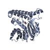 1kp9C 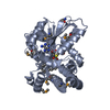 1kpgC 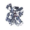 1kphC  1kpiC C: citing same article ( |
|---|---|
| Similar structure data | |
| Other databases |

-Links
 PDBj
PDBj


- Assembly
| Deposited unit | 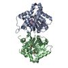
| ||||||||
|---|---|---|---|---|---|---|---|---|---|
| 1 | 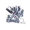
| ||||||||
| 2 | 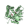
| ||||||||
| Unit cell |
| ||||||||
| Details | Biological assembly is a monomer |
-Components
| #1: Protein | Mass: 33070.676 Da / Num. of mol.: 2 Source method: isolated from a genetically manipulated source Source: (gene. exp.) Mycobacterium tuberculosis (bacteria) / Gene: pcaA / Plasmid: pET30b / Species (production host): Escherichia coli / Production host: References: UniProt: Q7D9R5, UniProt: P9WPB3*PLUS, cyclopropane-fatty-acyl-phospholipid synthase #2: Chemical |   Mass: 60.009 Da / Num. of mol.: 2 / Source method: obtained synthetically / Formula: CO3 Mass: 60.009 Da / Num. of mol.: 2 / Source method: obtained synthetically / Formula: CO3#3: Chemical |   Type: L-peptide linking / Mass: 384.411 Da / Num. of mol.: 2 / Source method: obtained synthetically / Formula: C14H20N6O5S Type: L-peptide linking / Mass: 384.411 Da / Num. of mol.: 2 / Source method: obtained synthetically / Formula: C14H20N6O5S#4: Water | ChemComp-HOH / |  Mass: 18.015 Da / Num. of mol.: 200 / Source method: isolated from a natural source / Formula: H2O Mass: 18.015 Da / Num. of mol.: 200 / Source method: isolated from a natural source / Formula: H2O |
|---|
-Experimental details
-Experiment
| Experiment | Method: X-RAY DIFFRACTION / Number of used crystals: 2 |
|---|
- Sample preparation
| Crystal | Density Matthews: 2 Å3/Da / Density % sol: 38.53 % |
|---|---|
| Crystal grow | Temperature: 292 K / Method: vapor diffusion, hanging drop / pH: 4.8 Details: PEG 6000, sodium acetate, n-octanoylsucrose, S-adenosyl-L-homocysteine, pH 4.8, VAPOR DIFFUSION, HANGING DROP, temperature 292K |
-Data collection
| Diffraction |
| ||||||||||||||||||
|---|---|---|---|---|---|---|---|---|---|---|---|---|---|---|---|---|---|---|---|
| Diffraction source |
| ||||||||||||||||||
| Detector |
| ||||||||||||||||||
| Radiation |
| ||||||||||||||||||
| Radiation wavelength |
| ||||||||||||||||||
| Reflection | Resolution: 2→30 Å / Num. all: 33417 / Num. obs: 33305 / % possible obs: 98 % / Observed criterion σ(F): 3 / Observed criterion σ(I): 3 / Biso Wilson estimate: 19.3 Å2 | ||||||||||||||||||
| Reflection shell | Resolution: 2→2.07 Å / % possible all: 93.4 |

- Processing
| Software |
| ||||||||||||||||||||||||||||||||||||
|---|---|---|---|---|---|---|---|---|---|---|---|---|---|---|---|---|---|---|---|---|---|---|---|---|---|---|---|---|---|---|---|---|---|---|---|---|---|
| Refinement | Method to determine structure: MAD / Resolution: 2→28.21 Å / Rfactor Rfree error: 0.007 / Data cutoff high absF: 450578.69 / Data cutoff low absF: 0 / Isotropic thermal model: RESTRAINED / Cross valid method: THROUGHOUT / σ(F): 3 / Stereochemistry target values: Engh & Huber Details: The electron density in the region of A175 to A180 was weak. The main chain was built in; however, there is some difficulty in fitting the side chain of Glu176, leading to a close contact ...Details: The electron density in the region of A175 to A180 was weak. The main chain was built in; however, there is some difficulty in fitting the side chain of Glu176, leading to a close contact with Lys180. The electron density corresponding to residues B176-B187 was not present. These residues were not built into the model.
| ||||||||||||||||||||||||||||||||||||
| Solvent computation | Solvent model: FLAT MODEL / Bsol: 37.1262 Å2 / ksol: 0.288251 e/Å3 | ||||||||||||||||||||||||||||||||||||
| Displacement parameters | Biso mean: 33.9 Å2
| ||||||||||||||||||||||||||||||||||||
| Refine analyze |
| ||||||||||||||||||||||||||||||||||||
| Refinement step | Cycle: LAST / Resolution: 2→28.21 Å
| ||||||||||||||||||||||||||||||||||||
| Refine LS restraints |
| ||||||||||||||||||||||||||||||||||||
| LS refinement shell | Resolution: 2→2.13 Å / Rfactor Rfree error: 0.021 / Total num. of bins used: 6
| ||||||||||||||||||||||||||||||||||||
| Xplor file |
|
