Type: DECTRIS EIGER X 16M / Detector: PIXEL / Date: May 24, 2016
Radiation
Protocol: SINGLE WAVELENGTH / Monochromatic (M) / Laue (L): M / Scattering type: x-ray
Radiation wavelength
Wavelength: 1.9 Å / Relative weight: 1
Reflection
Resolution: 2.6→44.84 Å / Num. obs: 19853 / % possible obs: 100 % / Redundancy: 17.91 % / CC1/2: 0.99 / Rrim(I) all: 0.4 / Net I/σ(I): 8.75
Reflection shell
Resolution: 2.6→2.67 Å / Redundancy: 12.33 % / Mean I/σ(I) obs: 1.87 / CC1/2: 0.25 / Rrim(I) all: 2.03 / % possible all: 99.9
-
Processing
Software
Name
Version
Classification
PHENIX
(1.12rc2_2821: ???)
refinement
XDS
datareduction
XSCALE
datascaling
SHELXCD
phasing
PHENIX
modelbuilding
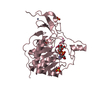
Refinement
Method to determine structure: SAD / Resolution: 2.6→44.455 Å / SU ML: 0.36 / Cross valid method: THROUGHOUT / σ(F): 1.32 / Phase error: 25.45 / Stereochemistry target values: ML
Rfactor
Num. reflection
% reflection
Rfree
0.2439
990
4.99 %
Rwork
0.2058
-
-
obs
0.2077
19853
99.93 %
Solvent computation
Shrinkage radii: 0.9 Å / VDW probe radii: 1.11 Å / Solvent model: FLAT BULK SOLVENT MODEL
Refinement step
Cycle: LAST / Resolution: 2.6→44.455 Å
Protein
Nucleic acid
Ligand
Solvent
Total
Num. atoms
2056
0
57
6
2119
Refine LS restraints
Refine-ID
Type
Dev ideal
Number
X-RAY DIFFRACTION
f_bond_d
0.006
2166
X-RAY DIFFRACTION
f_angle_d
0.865
2929
X-RAY DIFFRACTION
f_dihedral_angle_d
11.626
1251
X-RAY DIFFRACTION
f_chiral_restr
0.047
344
X-RAY DIFFRACTION
f_plane_restr
0.005
356
LS refinement shell
Resolution (Å)
Rfactor Rfree
Num. reflection Rfree
Rfactor Rwork
Num. reflection Rwork
Refine-ID
% reflection obs (%)
2.5999-2.7369
0.3397
143
0.2749
2716
X-RAY DIFFRACTION
100
2.7369-2.9084
0.3163
140
0.2576
2698
X-RAY DIFFRACTION
100
2.9084-3.1329
0.273
142
0.2286
2685
X-RAY DIFFRACTION
100
3.1329-3.4481
0.2635
140
0.2154
2689
X-RAY DIFFRACTION
100
3.4481-3.9467
0.2259
143
0.196
2682
X-RAY DIFFRACTION
100
3.9467-4.9714
0.2459
140
0.1823
2696
X-RAY DIFFRACTION
100
4.9714-44.4611
0.1944
142
0.1888
2697
X-RAY DIFFRACTION
100
+
About Yorodumi
-
News
-
Feb 9, 2022. New format data for meta-information of EMDB entries
New format data for meta-information of EMDB entries
Version 3 of the EMDB header file is now the official format.
The previous official version 1.9 will be removed from the archive.
In the structure databanks used in Yorodumi, some data are registered as the other names, "COVID-19 virus" and "2019-nCoV". Here are the details of the virus and the list of structure data.
Jan 31, 2019. EMDB accession codes are about to change! (news from PDBe EMDB page)
EMDB accession codes are about to change! (news from PDBe EMDB page)
The allocation of 4 digits for EMDB accession codes will soon come to an end. Whilst these codes will remain in use, new EMDB accession codes will include an additional digit and will expand incrementally as the available range of codes is exhausted. The current 4-digit format prefixed with “EMD-” (i.e. EMD-XXXX) will advance to a 5-digit format (i.e. EMD-XXXXX), and so on. It is currently estimated that the 4-digit codes will be depleted around Spring 2019, at which point the 5-digit format will come into force.
The EM Navigator/Yorodumi systems omit the EMD- prefix.
Related info.:Q: What is EMD? / ID/Accession-code notation in Yorodumi/EM Navigator
Yorodumi is a browser for structure data from EMDB, PDB, SASBDB, etc.
This page is also the successor to EM Navigator detail page, and also detail information page/front-end page for Omokage search.
The word "yorodu" (or yorozu) is an old Japanese word meaning "ten thousand". "mi" (miru) is to see.
Related info.:EMDB / PDB / SASBDB / Comparison of 3 databanks / Yorodumi Search / Aug 31, 2016. New EM Navigator & Yorodumi / Yorodumi Papers / Jmol/JSmol / Function and homology information / Changes in new EM Navigator and Yorodumi
 Movie
Movie Controller


 Open data
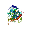
 Basic information
 Components
 Keywords
 Function and homology information
 X-RAY DIFFRACTION /
  Authors
 Ireland,
  Germany,
  France, 3items
  Citation
 Structure visualization
 Downloads & links
 Other downloads

 PDBj
PDBj


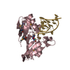
 Assembly
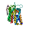

 Mass: 200.590 Da / Num. of mol.: 2 / Source method: obtained synthetically / Formula: Hg
 Mass: 122.143 Da / Num. of mol.: 1 / Source method: obtained synthetically / Formula: C4H12NO3 / Comment: pH buffer*YM
 Mass: 356.540 Da / Num. of mol.: 2 / Source method: obtained synthetically / Formula: C21H40O4
 Mass: 18.015 Da / Num. of mol.: 6 / Source method: isolated from a natural source / Formula: H2O
 Sample preparation
 / Beamline: X06SA / Wavelength: 1.9 Å
 Processing