 Movie
Movie Controller


+ Open data

- Basic information
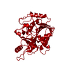
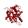
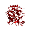
| Entry | Database: PDB / ID: 1pbn | ||||||
|---|---|---|---|---|---|---|---|
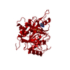
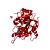
| Title | PURINE NUCLEOSIDE PHOSPHORYLASE | ||||||
 Components Components | PURINE NUCLEOSIDE PHOSPHORYLASE | ||||||
 Keywords Keywords | PENTOSYLTRANSFERASE / PURINE NUCLEOSIDE PHOSPHORYLASE | ||||||
| Function / homology |  Function and homology information Function and homology informationguanosine phosphorylase activity / purine-nucleoside phosphorylase activity / purine-nucleoside phosphorylase / purine ribonucleoside salvage / cytosol / cytoplasm Similarity search - Function | ||||||
| Biological species |  | ||||||
| Method |  X-RAY DIFFRACTION / Resolution: 2 Å X-RAY DIFFRACTION / Resolution: 2 Å | ||||||
 Authors Authors | Mao, C. / Ealick, S.E. | ||||||
 Citation Citation | Journal: Biochemistry / Year: 1998 Title: Calf spleen purine nucleoside phosphorylase complexed with substrates and substrate analogues. Authors: Mao, C. / Cook, W.J. / Zhou, M. / Federov, A.A. / Almo, S.C. / Ealick, S.E. | ||||||
| History |
|
- Structure visualization
| Structure viewer | Molecule: MolmilJmol/JSmol |
|---|
- Downloads & links
-Download
| PDBx/mmCIF format | 1pbn.cif.gz | 67.2 KB | Display | PDBx/mmCIF format |
|---|---|---|---|---|
| PDB format | pdb1pbn.ent.gz | 50.4 KB | Display | PDB format |
| PDBx/mmJSON format | 1pbn.json.gz | Tree view | PDBx/mmJSON format | |
| Others |  Other downloads Other downloads |
-Validation report
| Arichive directory | https://data.pdbj.org/pub/pdb/validation_reports/pb/1pbnftp://data.pdbj.org/pub/pdb/validation_reports/pb/1pbn | HTTPS FTP |
|---|
-Related structure data
| Related structure data |  1a9oC 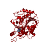 1a9pC 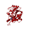 1a9qC 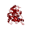 1a9rC 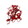 1a9sC 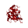 1a9tC C: citing same article ( |
|---|---|
| Similar structure data |


-Links
 PDBj
PDBj- Assembly
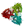
| Deposited unit | 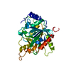
| ||||||||
|---|---|---|---|---|---|---|---|---|---|
| 1 | 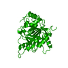
| ||||||||
| Unit cell |
| ||||||||
| Atom site foot note | 1: CIS PROLINE - PRO 198 | ||||||||
| Details | SYMMETRY THE CRYSTALLOGRAPHIC SYMMETRY TRANSFORMATIONS PRESENTED BELOW GENERATE THE SUBUNITS OF THE POLYMERIC MOLECULE. APPLIED TO RESIDUES: X 1 .. X 289 SYMMETRY1 1 0.000000 0.000000 -1.000000 94.20000 SYMMETRY2 1 1.000000 0.000000 0.000000 47.10000 SYMMETRY3 1 0.000000 -1.000000 0.000000 141.30000 APPLIED TO RESIDUES: X 1 .. X 289 SYMMETRY1 2 0.000000 1.000000 0.000000 -47.10000 SYMMETRY2 2 0.000000 0.000000 -1.000000 141.30000 SYMMETRY3 2 -1.000000 0.000000 0.000000 94.20000 |
-Components
| #1: Protein | Mass: 32008.420 Da / Num. of mol.: 1 / Source method: isolated from a natural source / Source: (natural) References: UniProt: P55859, purine-nucleoside phosphorylase |
|---|---|
| #2: Water | ChemComp-HOH /  Mass: 18.015 Da / Num. of mol.: 43 / Source method: isolated from a natural source / Formula: H2O Mass: 18.015 Da / Num. of mol.: 43 / Source method: isolated from a natural source / Formula: H2O |
-Experimental details
-Experiment
| Experiment | Method: X-RAY DIFFRACTION / Number of used crystals: 1 |
|---|
- Sample preparation
| Crystal | Density Matthews: 2.18 Å3/Da / Density % sol: 43.46 % | ||||||||||||||||||||||||
|---|---|---|---|---|---|---|---|---|---|---|---|---|---|---|---|---|---|---|---|---|---|---|---|---|---|
| Crystal | *PLUS Density % sol: 65 % | ||||||||||||||||||||||||
| Crystal grow | *PLUS Method: vapor diffusion, hanging drop / PH range low: 8.2 / PH range high: 7.8 | ||||||||||||||||||||||||
| Components of the solutions | *PLUS
|
-Data collection
| Diffraction source | Wavelength: 1.5418 Å |
|---|---|
| Detector | Type: XUONG-HAMLIN MULTIWIRE / Detector: AREA DETECTOR |
| Radiation | Monochromatic (M) / Laue (L): M / Scattering type: x-ray |
| Radiation wavelength | Wavelength: 1.5418 Å / Relative weight: 1 |
| Reflection | Num. obs: 17293 / % possible obs: 80 % / Observed criterion σ(I): 2 / Redundancy: 2.9 % / Rmerge(I) obs: 0.13 |
- Processing
| Software |
| ||||||||||||||||||||||||||||||||||||||||||||||||||||||||||||
|---|---|---|---|---|---|---|---|---|---|---|---|---|---|---|---|---|---|---|---|---|---|---|---|---|---|---|---|---|---|---|---|---|---|---|---|---|---|---|---|---|---|---|---|---|---|---|---|---|---|---|---|---|---|---|---|---|---|---|---|---|---|
| Refinement | Resolution: 2→15 Å / σ(F): 2 /
| ||||||||||||||||||||||||||||||||||||||||||||||||||||||||||||
| Refinement step | Cycle: LAST / Resolution: 2→15 Å
| ||||||||||||||||||||||||||||||||||||||||||||||||||||||||||||
| Refine LS restraints |
| ||||||||||||||||||||||||||||||||||||||||||||||||||||||||||||
| Software | *PLUS Name: X-PLOR / Classification: refinement | ||||||||||||||||||||||||||||||||||||||||||||||||||||||||||||
| Refinement | *PLUS | ||||||||||||||||||||||||||||||||||||||||||||||||||||||||||||
| Solvent computation | *PLUS | ||||||||||||||||||||||||||||||||||||||||||||||||||||||||||||
| Displacement parameters | *PLUS | ||||||||||||||||||||||||||||||||||||||||||||||||||||||||||||
| Refine LS restraints | *PLUS Type: x_improper_angle_deg / Dev ideal: 1.18 |
