 Movie
Movie Controller

[English] 日本語
 Yorodumi
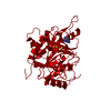
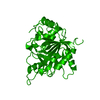
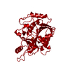
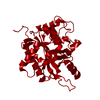
- PDB-1ula: APPLICATION OF CRYSTALLOGRAPHIC AND MODELING METHODS IN THE DESIG... -
+ Open data

- Basic information
| Entry | Database: PDB / ID: 1ula | |||||||||
|---|---|---|---|---|---|---|---|---|---|---|
| Title | APPLICATION OF CRYSTALLOGRAPHIC AND MODELING METHODS IN THE DESIGN OF PURINE NUCLEOSIDE PHOSPHORYLASE INHIBITORS | |||||||||
 Components Components | PURINE NUCLEOSIDE PHOSPHORYLASE | |||||||||
 Keywords Keywords | PENTOSYLTRANSFERASE | |||||||||
| Function / homology |  Function and homology information Function and homology informationnicotinamide riboside catabolic process / Defective PNP disrupts phosphorolysis of (deoxy)guanosine and (deoxy)inosine / purine-containing compound salvage / deoxyinosine catabolic process / purine nucleobase binding / deoxyadenosine catabolic process / nucleotide biosynthetic process / inosine catabolic process / dAMP catabolic process / urate biosynthetic process ...nicotinamide riboside catabolic process / Defective PNP disrupts phosphorolysis of (deoxy)guanosine and (deoxy)inosine / purine-containing compound salvage / deoxyinosine catabolic process / purine nucleobase binding / deoxyadenosine catabolic process / nucleotide biosynthetic process / inosine catabolic process / dAMP catabolic process / urate biosynthetic process / IMP catabolic process / Ribavirin ADME / guanosine phosphorylase activity / nucleoside binding / allantoin metabolic process / Purine salvage / Purine catabolism / purine-nucleoside phosphorylase / purine-nucleoside phosphorylase activity / positive regulation of alpha-beta T cell differentiation / purine ribonucleoside salvage / nucleobase-containing compound metabolic process / phosphate ion binding / positive regulation of interleukin-2 production / positive regulation of T cell proliferation / secretory granule lumen / ficolin-1-rich granule lumen / immune response / response to xenobiotic stimulus / Neutrophil degranulation / extracellular exosome / extracellular region / identical protein binding / cytoplasm / cytosol Similarity search - Function | |||||||||
| Biological species |  Homo sapiens (human) Homo sapiens (human) | |||||||||
| Method |  X-RAY DIFFRACTION / Resolution: 2.75 Å X-RAY DIFFRACTION / Resolution: 2.75 Å | |||||||||
 Authors Authors | Ealick, S.E. / Rule, S.A. / Carter, D.C. / Greenhough, T.J. / Babu, Y.S. / Cook, W.J. / Habash, J. / Helliwell, J.R. / Stoeckler, J.D. / Parksjunior, R.E. ...Ealick, S.E. / Rule, S.A. / Carter, D.C. / Greenhough, T.J. / Babu, Y.S. / Cook, W.J. / Habash, J. / Helliwell, J.R. / Stoeckler, J.D. / Parksjunior, R.E. / Chen, S.-F. / Bugg, C.E. | |||||||||
 Citation Citation | Journal: Proc.Natl.Acad.Sci.USA / Year: 1991 Title: Application of crystallographic and modeling methods in the design of purine nucleoside phosphorylase inhibitors. Authors: Ealick, S.E. / Babu, Y.S. / Bugg, C.E. / Erion, M.D. / Guida, W.C. / Montgomery, J.A. / Secrist 3rd., J.A. #1: Journal: J.Biol.Chem. / Year: 1990Title: Three-Dimensional Structure of Human Erythrocytic Purine Nucleoside Phosphorylase at 3.2 Angstroms Resolution Authors: Ealick, S.E. / Rule, S.A. / Carter, D.C. / Greenhough, T.J. / Babu, Y.S. / Cook, W.J. / Habash, J. / Helliwell, J.R. / Stoeckler, J.D. / Parksjunior, R.E. / Chen, S.-F. / Bugg, C.E. #2: Journal: J.Biol.Chem. / Year: 1981Title: Crystallization and Preliminary X-Ray Investigation of Human Erythrocyte Purine Nucleoside Phosphorylase Authors: Cook, W.J. / Ealick, S.E. / Bugg, C.E. / Stoeckler, J.D. / Parksjunior, R.E. | |||||||||
| History |
|
- Structure visualization
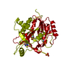
| Structure viewer | Molecule: MolmilJmol/JSmol |
|---|
- Downloads & links
-Download
| PDBx/mmCIF format | 1ula.cif.gz | 67.7 KB | Display | PDBx/mmCIF format |
|---|---|---|---|---|
| PDB format | pdb1ula.ent.gz | 51 KB | Display | PDB format |
| PDBx/mmJSON format | 1ula.json.gz | Tree view | PDBx/mmJSON format | |
| Others |  Other downloads Other downloads |
-Validation report
| Arichive directory | https://data.pdbj.org/pub/pdb/validation_reports/ul/1ulaftp://data.pdbj.org/pub/pdb/validation_reports/ul/1ula | HTTPS FTP |
|---|
-Related structure data


-Links
 PDBj
PDBj
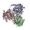
- Assembly
| Deposited unit | 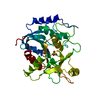
| ||||||||
|---|---|---|---|---|---|---|---|---|---|
| 1 | 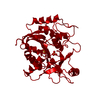
| ||||||||
| Unit cell |
|
-Components
| #1: Protein | Mass: 32184.877 Da / Num. of mol.: 1 Source method: isolated from a genetically manipulated source Source: (gene. exp.) Homo sapiens (human)References: UniProt: P00491, purine-nucleoside phosphorylase |
|---|---|
| #2: Chemical |   Mass: 96.063 Da / Num. of mol.: 2 / Source method: obtained synthetically / Formula: SO4 Mass: 96.063 Da / Num. of mol.: 2 / Source method: obtained synthetically / Formula: SO4 |
-Experimental details
-Experiment
| Experiment | Method: X-RAY DIFFRACTION |
|---|
- Sample preparation
| Crystal | Density Matthews: 5.04 Å3/Da / Density % sol: 75.61 % |
|---|---|
| Crystal grow | *PLUS Method: unknown |
- Processing
| Software | Name: PROLSQ / Classification: refinement | ||||||||||||
|---|---|---|---|---|---|---|---|---|---|---|---|---|---|
| Refinement | Rfactor obs: 0.202 / Highest resolution: 2.75 Å | ||||||||||||
| Refinement step | Cycle: LAST / Highest resolution: 2.75 Å
| ||||||||||||
| Refine LS restraints |
|
