 Movie
Movie Controller


+ Open data

- Basic information
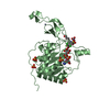
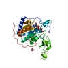
| Entry | Database: PDB / ID: 3pki | ||||||
|---|---|---|---|---|---|---|---|
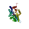
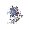
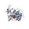
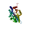
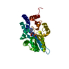
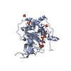
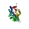
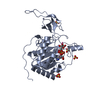
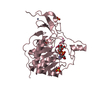
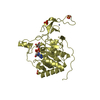
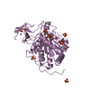
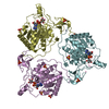
| Title | Human SIRT6 crystal structure in complex with ADP ribose | ||||||
 Components Components | NAD-dependent deacetylase sirtuin-6 | ||||||
 Keywords Keywords | HYDROLASE / SIRT6 / ADP ribose / STRUCTURAL GENOMICS / STRUCTURAL GENOMICS CONSORTIUM / SGC | ||||||
| Function / homology |  Function and homology information Function and homology informationhistone H3K56 deacetylase activity, NAD-dependent / ketone biosynthetic process / histone H3K18 deacetylase activity, NAD-dependent / histone H3K9 deacetylase activity, hydrolytic mechanism / histone H3K9 deacetylase activity, NAD-dependent / protein delipidation / NAD+-protein-lysine ADP-ribosyltransferase activity / chromosome, subtelomeric region / regulation of lipid catabolic process / positive regulation of protein localization to chromatin ...histone H3K56 deacetylase activity, NAD-dependent / ketone biosynthetic process / histone H3K18 deacetylase activity, NAD-dependent / histone H3K9 deacetylase activity, hydrolytic mechanism / histone H3K9 deacetylase activity, NAD-dependent / protein delipidation / NAD+-protein-lysine ADP-ribosyltransferase activity / chromosome, subtelomeric region / regulation of lipid catabolic process / positive regulation of protein localization to chromatin / NAD+-protein-arginine ADP-ribosyltransferase activity / NAD-dependent protein demyristoylase activity / NAD-dependent protein depalmitoylase activity / DNA damage sensor activity / positive regulation of stem cell differentiation / negative regulation of D-glucose import across plasma membrane / positive regulation of chondrocyte proliferation / transposable element silencing / protein acetyllysine N-acetyltransferase / protein deacetylation / NAD-dependent protein lysine deacetylase activity / histone deacetylase activity, NAD-dependent / pericentric heterochromatin formation / positive regulation of blood vessel branching / cardiac muscle cell differentiation / protein localization to site of double-strand break / positive regulation of vascular endothelial cell proliferation / negative regulation of glycolytic process / TORC2 complex binding / negative regulation of protein import into nucleus / regulation of protein secretion / regulation of double-strand break repair via homologous recombination / positive regulation of stem cell proliferation / positive regulation of double-strand break repair / negative regulation of gene expression, epigenetic / lncRNA binding / NAD+-protein mono-ADP-ribosyltransferase activity / DNA repair-dependent chromatin remodeling / positive regulation of stem cell population maintenance / positive regulation of telomere maintenance / negative regulation of cellular senescence / Transferases; Glycosyltransferases; Pentosyltransferases / negative regulation of transcription elongation by RNA polymerase II / regulation of lipid metabolic process / NAD+ poly-ADP-ribosyltransferase activity / NAD+ binding / negative regulation of gluconeogenesis / subtelomeric heterochromatin formation / positive regulation of fat cell differentiation / response to UV / pericentric heterochromatin / nucleosome binding / regulation of protein localization to plasma membrane / site of DNA damage / negative regulation of protein localization to chromatin / nucleotidyltransferase activity / Transferases; Acyltransferases; Transferring groups other than aminoacyl groups / positive regulation of protein export from nucleus / determination of adult lifespan / circadian regulation of gene expression / chromatin DNA binding / regulation of circadian rhythm / base-excision repair / protein destabilization / Pre-NOTCH Transcription and Translation / positive regulation of fibroblast proliferation / positive regulation of insulin secretion / protein import into nucleus / transcription corepressor activity / double-strand break repair / positive regulation of proteasomal ubiquitin-dependent protein catabolic process / glucose homeostasis / positive regulation of cold-induced thermogenesis / site of double-strand break / Processing of DNA double-strand break ends / damaged DNA binding / chromatin remodeling / negative regulation of cell population proliferation / chromatin binding / chromatin / negative regulation of transcription by RNA polymerase II / endoplasmic reticulum / protein homodimerization activity / DNA binding / zinc ion binding / nucleoplasm / nucleus Similarity search - Function | ||||||
| Biological species |  Homo sapiens (human) Homo sapiens (human) | ||||||
| Method |  X-RAY DIFFRACTION / SYNCHROTRON / MOLECULAR REPLACEMENT / Resolution: 2.04 Å X-RAY DIFFRACTION / SYNCHROTRON / MOLECULAR REPLACEMENT / Resolution: 2.04 Å | ||||||
 Authors Authors | Pan, P.W. / Dong, A. / Qiu, W. / Loppnau, P. / Wang, J. / Ravichandran, M. / Bochkarev, A. / Bountra, C. / Weigelt, J. / Arrowsmith, C.H. ...Pan, P.W. / Dong, A. / Qiu, W. / Loppnau, P. / Wang, J. / Ravichandran, M. / Bochkarev, A. / Bountra, C. / Weigelt, J. / Arrowsmith, C.H. / Min, J. / Edwards, A.M. / Structural Genomics Consortium (SGC) | ||||||
 Citation Citation | Journal: J.Biol.Chem. / Year: 2011 Title: Structure and biochemical functions of SIRT6. Authors: Pan, P.W. / Feldman, J.L. / Devries, M.K. / Dong, A. / Edwards, A.M. / Denu, J.M. | ||||||
| History |
|
- Structure visualization
| Structure viewer | Molecule: MolmilJmol/JSmol |
|---|
- Downloads & links
-Download
| PDBx/mmCIF format | 3pki.cif.gz | 367.9 KB | Display | PDBx/mmCIF format |
|---|---|---|---|---|
| PDB format | pdb3pki.ent.gz | 293.5 KB | Display | PDB format |
| PDBx/mmJSON format | 3pki.json.gz | Tree view | PDBx/mmJSON format | |
| Others |  Other downloads Other downloads |
-Validation report
| Arichive directory | https://data.pdbj.org/pub/pdb/validation_reports/pk/3pkiftp://data.pdbj.org/pub/pdb/validation_reports/pk/3pki | HTTPS FTP |
|---|
-Related structure data
| Related structure data | 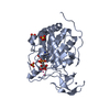 3k35SC 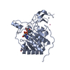 3pkjC C: citing same article ( S: Starting model for refinement |
|---|---|
| Similar structure data |

-Links
 PDBj
PDBj

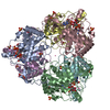
- Assembly


-Components
-Protein , 1 types, 6 molecules ABCDEF
| #1: Protein | Mass: 39106.707 Da / Num. of mol.: 6 / Mutation: K265E Source method: isolated from a genetically manipulated source Source: (gene. exp.) Homo sapiens (human) / Gene: SIRT6, SIR2L6 / Plasmid: PET28-LIC / Production host:  References: UniProt: Q8N6T7, Hydrolases; Acting on carbon-nitrogen bonds, other than peptide bonds; In linear amides |
|---|
-Non-polymers , 5 types, 1000 molecules 


| #2: Chemical | ChemComp-UNX /  Num. of mol.: 26 / Source method: obtained synthetically Num. of mol.: 26 / Source method: obtained synthetically#3: Chemical | ChemComp-ZN /  Mass: 65.409 Da / Num. of mol.: 6 / Source method: obtained synthetically / Formula: Zn Mass: 65.409 Da / Num. of mol.: 6 / Source method: obtained synthetically / Formula: Zn#4: Chemical | ChemComp-AR6 / [(  Mass: 559.316 Da / Num. of mol.: 6 / Source method: obtained synthetically / Formula: C15H23N5O14P2 Mass: 559.316 Da / Num. of mol.: 6 / Source method: obtained synthetically / Formula: C15H23N5O14P2#5: Chemical | ChemComp-SO4 /  Mass: 96.063 Da / Num. of mol.: 23 / Source method: obtained synthetically / Formula: SO4 Mass: 96.063 Da / Num. of mol.: 23 / Source method: obtained synthetically / Formula: SO4#6: Water | ChemComp-HOH / | Mass: 18.015 Da / Num. of mol.: 939 / Source method: isolated from a natural source / Formula: H2O |
|---|
-Experimental details
-Experiment
| Experiment | Method: X-RAY DIFFRACTION / Number of used crystals: 1 |
|---|
- Sample preparation
| Crystal | Density Matthews: 2.04 Å3/Da / Density % sol: 39.7 % |
|---|---|
| Crystal grow | Temperature: 297 K / pH: 5.8 Details: 2M NH4SO4, 2%PEG400, 0.1M BIS-TRIS pH5.8, temperature 297K |
-Data collection
| Diffraction | Mean temperature: 100 K |
|---|---|
| Diffraction source | Source: SYNCHROTRON / Site: APS  / Beamline: 19-ID / Wavelength: 0.97945 Å / Beamline: 19-ID / Wavelength: 0.97945 Å |
| Detector | Type: ADSC QUANTUM 315 / Detector: CCD / Date: Oct 14, 2009 |
| Radiation | Protocol: SINGLE WAVELENGTH / Monochromatic (M) / Laue (L): M / Scattering type: x-ray |
| Radiation wavelength | Wavelength: 0.97945 Å / Relative weight: 1 |
| Reflection | Resolution: 2.03→50 Å / Num. all: 118501 / Num. obs: 118501 / % possible obs: 98.2 % / Observed criterion σ(F): 0 / Observed criterion σ(I): 0 / Redundancy: 4.5 % / Biso Wilson estimate: 29.47 Å2 / Rmerge(I) obs: 0.119 / Rsym value: 0.119 / Net I/σ(I): 14.6 |
| Reflection shell | Resolution: 2.03→2.07 Å / Redundancy: 4.1 % / Rmerge(I) obs: 0.956 / Mean I/σ(I) obs: 1.7 / Num. unique all: 5734 / Rsym value: 0.956 / % possible all: 97 |
- Processing
| Software |
| |||||||||||||||||||||||||||||||||||||||||||||||||||||||||||||||||||||||||||||||||||||||||||||||
|---|---|---|---|---|---|---|---|---|---|---|---|---|---|---|---|---|---|---|---|---|---|---|---|---|---|---|---|---|---|---|---|---|---|---|---|---|---|---|---|---|---|---|---|---|---|---|---|---|---|---|---|---|---|---|---|---|---|---|---|---|---|---|---|---|---|---|---|---|---|---|---|---|---|---|---|---|---|---|---|---|---|---|---|---|---|---|---|---|---|---|---|---|---|---|---|---|
| Refinement | Method to determine structure: MOLECULAR REPLACEMENT Starting model: 3K35 Resolution: 2.04→29.87 Å / Cor.coef. Fo:Fc: 0.9385 / Cor.coef. Fo:Fc free: 0.9221 / Cross valid method: THROUGHOUT / σ(F): 0 / σ(I): 0 / Stereochemistry target values: Engh & Huber
| |||||||||||||||||||||||||||||||||||||||||||||||||||||||||||||||||||||||||||||||||||||||||||||||
| Displacement parameters | Biso mean: 33.39 Å2
| |||||||||||||||||||||||||||||||||||||||||||||||||||||||||||||||||||||||||||||||||||||||||||||||
| Refine analyze | Luzzati coordinate error obs: 0.242 Å | |||||||||||||||||||||||||||||||||||||||||||||||||||||||||||||||||||||||||||||||||||||||||||||||
| Refinement step | Cycle: LAST / Resolution: 2.04→29.87 Å
| |||||||||||||||||||||||||||||||||||||||||||||||||||||||||||||||||||||||||||||||||||||||||||||||
| Refine LS restraints |
| |||||||||||||||||||||||||||||||||||||||||||||||||||||||||||||||||||||||||||||||||||||||||||||||
| LS refinement shell | Resolution: 2.04→2.09 Å / Total num. of bins used: 20
|
