[English] 日本語
 Yorodumi
- PDB-3ckx: Crystal structure of sterile 20-like kinase 3 (MST3, STK24) in co... -
+ Open data
- Basic information
| Entry | Database: PDB / ID: 3ckx | ||||||
|---|---|---|---|---|---|---|---|
| Title | Crystal structure of sterile 20-like kinase 3 (MST3, STK24) in complex with staurosporine | ||||||
 Components Components | Serine/threonine-protein kinase 24 | ||||||
 Keywords Keywords | TRANSFERASE / STRUCTURAL GENOMICS / PSI-2 / PROTEIN STRUCTURE INITIATIVE / New York SGX Research Center for Structural Genomics / NYSGXRC / PROTEIN KINASE / MST3 / STK24 / STERILE 20-LIKE KINASE / STAUROSPORINE / Alternative splicing / ATP-binding / Cytoplasm / Nucleotide-binding / Phosphoprotein / Serine/threonine-protein kinase | ||||||
| Function / homology |  Function and homology information Function and homology informationApoptotic execution phase / FAR/SIN/STRIPAK complex / regulation of axon regeneration / intrinsic apoptotic signaling pathway in response to oxidative stress / execution phase of apoptosis / Apoptotic cleavage of cellular proteins / negative regulation of cell migration / cellular response to starvation / protein autophosphorylation / cellular response to oxidative stress ...Apoptotic execution phase / FAR/SIN/STRIPAK complex / regulation of axon regeneration / intrinsic apoptotic signaling pathway in response to oxidative stress / execution phase of apoptosis / Apoptotic cleavage of cellular proteins / negative regulation of cell migration / cellular response to starvation / protein autophosphorylation / cellular response to oxidative stress / protein phosphorylation / protein kinase activity / non-specific serine/threonine protein kinase / intracellular signal transduction / cadherin binding / protein serine kinase activity / protein serine/threonine kinase activity / nucleolus / Golgi apparatus / signal transduction / extracellular exosome / nucleoplasm / ATP binding / metal ion binding / nucleus / membrane / cytosol / cytoplasm Similarity search - Function | ||||||
| Biological species |  Homo sapiens (human) Homo sapiens (human) | ||||||
| Method |  X-RAY DIFFRACTION / X-RAY DIFFRACTION /  SYNCHROTRON / SYNCHROTRON /  MOLECULAR REPLACEMENT / Resolution: 2.7 Å MOLECULAR REPLACEMENT / Resolution: 2.7 Å | ||||||
 Authors Authors | Antonysamy, S.S. / Burley, S.K. / Buchanan, S. / Chau, F. / Feil, I. / Wu, L. / Sauder, J.M. / New York SGX Research Center for Structural Genomics (NYSGXRC) | ||||||
 Citation Citation |  Journal: To be Published Journal: To be PublishedTitle: Crystal structure of sterile 20-like kinase 3 (MST3, STK24) in complex with staurosporine. Authors: Antonysamy, S.S. / Burley, S.K. / Buchanan, S. / Chau, F. / Feil, I. / Wu, L. / Sauder, J.M. | ||||||
| History |
|
- Structure visualization
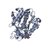
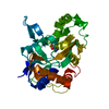
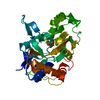
| Structure viewer | Molecule:  Molmil Molmil Jmol/JSmol Jmol/JSmol |
|---|
- Downloads & links
- Download
| PDBx/mmCIF format |  3ckx.cif.gz 3ckx.cif.gz | 69.4 KB | Display |  PDBx/mmCIF format PDBx/mmCIF format |
|---|---|---|---|---|
| PDB format |  pdb3ckx.ent.gz pdb3ckx.ent.gz | 50 KB | Display |  PDB format PDB format |
| PDBx/mmJSON format |  3ckx.json.gz 3ckx.json.gz | Tree view |  PDBx/mmJSON format PDBx/mmJSON format | |
| Others |  Other downloads Other downloads |
-Validation report
| Arichive directory |  https://data.pdbj.org/pub/pdb/validation_reports/ck/3ckx https://data.pdbj.org/pub/pdb/validation_reports/ck/3ckx ftp://data.pdbj.org/pub/pdb/validation_reports/ck/3ckx ftp://data.pdbj.org/pub/pdb/validation_reports/ck/3ckx | HTTPS FTP |
|---|
-Related structure data
| Related structure data |  3ckwS S: Starting model for refinement |
|---|---|
| Similar structure data | |
| Other databases |
- Links
Links
- Assembly
| Deposited unit | 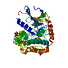
| ||||||||
|---|---|---|---|---|---|---|---|---|---|
| 1 |
| ||||||||
| Unit cell |
|
- Components
| #1: Protein | Mass: 34653.211 Da / Num. of mol.: 1 / Fragment: Residues 31-323 Source method: isolated from a genetically manipulated source Source: (gene. exp.)  Homo sapiens (human) / Gene: STK24, MST3, STK3 / Plasmid: BC-pSGX3(BC) / Production host: Homo sapiens (human) / Gene: STK24, MST3, STK3 / Plasmid: BC-pSGX3(BC) / Production host:  References: UniProt: Q9Y6E0, non-specific serine/threonine protein kinase |
|---|---|
| #2: Chemical | ChemComp-STU / |
| #3: Water | ChemComp-HOH / |
| Has protein modification | Y |
| Sequence details | THE TARGETDB ID NYSGXRC-5632A PROVIDED BY AUTHORS FOR THIS PROTEIN DID NOT EXIST IN TARGET DATABASE ...THE TARGETDB ID NYSGXRC-5632A PROVIDED BY AUTHORS FOR THIS PROTEIN DID NOT EXIST IN TARGET DATABASE AT THE TIME OF DEPOSITION |
-Experimental details
-Experiment
| Experiment | Method:  X-RAY DIFFRACTION / Number of used crystals: 1 X-RAY DIFFRACTION / Number of used crystals: 1 |
|---|
- Sample preparation
| Crystal | Density Matthews: 2.08 Å3/Da / Density % sol: 40.84 % |
|---|---|
| Crystal grow | Temperature: 292 K / Method: vapor diffusion / pH: 8.5 Details: 22% PEG 4000, 100 mM Tris-HCl, 200 mM Sodium acetate, pH 8.5, VAPOR DIFFUSION, temperature 292K |
-Data collection
| Diffraction | Mean temperature: 100 K |
|---|---|
| Diffraction source | Source:  SYNCHROTRON / Site: SYNCHROTRON / Site:  APS APS  / Beamline: 31-ID / Wavelength: 0.9795 Å / Beamline: 31-ID / Wavelength: 0.9795 Å |
| Detector | Type: MAR CCD 165 mm / Detector: CCD / Date: Jan 9, 2001 |
| Radiation | Monochromator: DIAMOND / Protocol: SINGLE WAVELENGTH / Monochromatic (M) / Laue (L): M / Scattering type: x-ray |
| Radiation wavelength | Wavelength: 0.9795 Å / Relative weight: 1 |
| Reflection | Resolution: 2.7→50 Å / Num. obs: 7930 |
- Processing
| Software |
| ||||||||||||||||||||||||||||
|---|---|---|---|---|---|---|---|---|---|---|---|---|---|---|---|---|---|---|---|---|---|---|---|---|---|---|---|---|---|
| Refinement | Method to determine structure:  MOLECULAR REPLACEMENT MOLECULAR REPLACEMENTStarting model: PDB entry 3CKW Resolution: 2.7→24.77 Å
| ||||||||||||||||||||||||||||
| Displacement parameters | Biso mean: 56.855 Å2 | ||||||||||||||||||||||||||||
| Refinement step | Cycle: LAST / Resolution: 2.7→24.77 Å
|
 Movie
Movie Controller





 PDBj
PDBj
