| Entry | Database: PDB / ID: 2jks
|
|---|
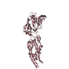
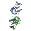
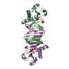
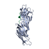
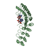
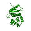
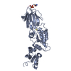
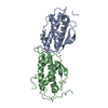
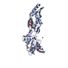
| Title | Crystal structure of the the bradyzoite specific antigen BSR4 from toxoplasma gondii. |
|---|
 Components Components | BRADYZOITE SURFACE ANTIGEN BSR4 |
|---|
 Keywords Keywords | IMMUNE SYSTEM |
|---|
| Function / homology |  Function and homology information Function and homology information
SRS domain / Protozoan surface antigen, SAG1 family / SRS domain / SRS domain / SRS domain superfamily / Immunoglobulin-like / Sandwich / Mainly BetaSimilarity search - Domain/homology |
|---|
| Biological species |   TOXOPLASMA GONDII (eukaryote) TOXOPLASMA GONDII (eukaryote) |
|---|
| Method |  X-RAY DIFFRACTION / MOLECULAR REPLACEMENT / Resolution: 1.9 Å X-RAY DIFFRACTION / MOLECULAR REPLACEMENT / Resolution: 1.9 Å |
|---|
 Authors Authors | Boulanger, M.J. / Grigg, M.E. / Bruic, E. / Grujic, O. |
|---|
 Citation Citation | Journal: J.Biol.Chem. / Year: 2009
Title: Structural Characterization of the Bradyzoite Surface Antigen (Bsr4) from Toxoplasma Gondii, a Unique Addition to the Surface Antigen Glycoprotein 1-Related Superfamily.
Authors: Crawford, J. / Grujic, O. / Bruic, E. / Czjzek, M. / Grigg, M.E. / Boulanger, M.J. |
|---|
| History | | Deposition | Aug 29, 2008 | Deposition site: PDBE / Processing site: PDBE |
|---|
| Revision 1.0 | Feb 3, 2009 | Provider: repository / Type: Initial release |
|---|
| Revision 1.1 | Jul 13, 2011 | Group: Advisory / Version format compliance |
|---|
| Revision 1.2 | Jul 5, 2017 | Group: Data collection / Category: diffrn_source / Item: _diffrn_source.type |
|---|
| Revision 1.3 | Dec 13, 2023 | Group: Data collection / Database references ...Data collection / Database references / Derived calculations / Other / Refinement description
Category: chem_comp_atom / chem_comp_bond ...chem_comp_atom / chem_comp_bond / database_2 / pdbx_database_status / pdbx_initial_refinement_model / pdbx_struct_conn_angle / struct_conn / struct_site
Item: _database_2.pdbx_DOI / _database_2.pdbx_database_accession ..._database_2.pdbx_DOI / _database_2.pdbx_database_accession / _pdbx_database_status.status_code_sf / _pdbx_struct_conn_angle.ptnr1_auth_comp_id / _pdbx_struct_conn_angle.ptnr1_auth_seq_id / _pdbx_struct_conn_angle.ptnr1_label_asym_id / _pdbx_struct_conn_angle.ptnr1_label_atom_id / _pdbx_struct_conn_angle.ptnr1_label_comp_id / _pdbx_struct_conn_angle.ptnr1_label_seq_id / _pdbx_struct_conn_angle.ptnr1_symmetry / _pdbx_struct_conn_angle.ptnr2_auth_seq_id / _pdbx_struct_conn_angle.ptnr2_label_asym_id / _pdbx_struct_conn_angle.ptnr3_auth_comp_id / _pdbx_struct_conn_angle.ptnr3_auth_seq_id / _pdbx_struct_conn_angle.ptnr3_label_asym_id / _pdbx_struct_conn_angle.ptnr3_label_atom_id / _pdbx_struct_conn_angle.ptnr3_label_comp_id / _pdbx_struct_conn_angle.ptnr3_label_seq_id / _pdbx_struct_conn_angle.ptnr3_symmetry / _pdbx_struct_conn_angle.value / _struct_conn.pdbx_dist_value / _struct_conn.ptnr1_auth_comp_id / _struct_conn.ptnr1_auth_seq_id / _struct_conn.ptnr1_label_asym_id / _struct_conn.ptnr1_label_atom_id / _struct_conn.ptnr1_label_comp_id / _struct_conn.ptnr1_label_seq_id / _struct_conn.ptnr1_symmetry / _struct_conn.ptnr2_auth_comp_id / _struct_conn.ptnr2_auth_seq_id / _struct_conn.ptnr2_label_asym_id / _struct_conn.ptnr2_label_atom_id / _struct_conn.ptnr2_label_comp_id / _struct_conn.ptnr2_label_seq_id / _struct_conn.ptnr2_symmetry / _struct_site.pdbx_auth_asym_id / _struct_site.pdbx_auth_comp_id / _struct_site.pdbx_auth_seq_id |
|---|
| Revision 1.4 | Oct 23, 2024 | Group: Structure summary / Category: pdbx_entry_details / pdbx_modification_feature |
|---|
|
|---|
 Movie
Movie Controller

 Yorodumi
 Open data
 Basic information
 Structure visualization
 Downloads & links
 Other downloads

 PDBj
PDBj Assembly
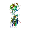

 SPODOPTERA FRUGIPERDA (fall armyworm) / Strain (production host): HI5 / References: UniProt: Q95NL6
 Mass: 65.409 Da / Num. of mol.: 8 / Source method: obtained synthetically / Formula: Zn
 Mass: 18.015 Da / Num. of mol.: 279 / Source method: isolated from a natural source / Formula: H2O
 Sample preparation
 Processing