 Movie
Movie Controller

[English] 日本語
 Yorodumi
- PDB-3if0: Crystal Structure of the Nanoarchaeum equitans tRNA splicing endo... -
+ Open data

- Basic information
| Entry | Database: PDB / ID: 3if0 | ||||||
|---|---|---|---|---|---|---|---|
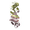
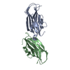
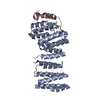
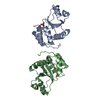
| Title | Crystal Structure of the Nanoarchaeum equitans tRNA splicing endonuclease structural subunit | ||||||
 Components Components | NEQ261 | ||||||
 Keywords Keywords | HYDROLASE / protein | ||||||
| Function / homology |  Function and homology information Function and homology informationtRNA-intron lyase activity / tRNA splicing, via endonucleolytic cleavage and ligation Similarity search - Function | ||||||
| Biological species |   Nanoarchaeum equitans (archaea) Nanoarchaeum equitans (archaea) | ||||||
| Method |  X-RAY DIFFRACTION / SYNCHROTRON / MOLECULAR REPLACEMENT / molecular replacement / Resolution: 2.2 Å X-RAY DIFFRACTION / SYNCHROTRON / MOLECULAR REPLACEMENT / molecular replacement / Resolution: 2.2 Å | ||||||
 Authors Authors | Mitchell, M. / Li, H. | ||||||
 Citation Citation | Journal: Nucleic Acids Res. / Year: 2009 Title: Crystal structure and assembly of the functional Nanoarchaeum equitans tRNA splicing endonuclease. Authors: Mitchell, M. / Xue, S. / Erdman, R. / Randau, L. / Soll, D. / Li, H. | ||||||
| History |
|
- Structure visualization
| Structure viewer | Molecule: MolmilJmol/JSmol |
|---|
- Downloads & links
-Download
| PDBx/mmCIF format | 3if0.cif.gz | 43.3 KB | Display | PDBx/mmCIF format |
|---|---|---|---|---|
| PDB format | pdb3if0.ent.gz | 30.8 KB | Display | PDB format |
| PDBx/mmJSON format | 3if0.json.gz | Tree view | PDBx/mmJSON format | |
| Others |  Other downloads Other downloads |
-Validation report
| Arichive directory | https://data.pdbj.org/pub/pdb/validation_reports/if/3if0ftp://data.pdbj.org/pub/pdb/validation_reports/if/3if0 | HTTPS FTP |
|---|
-Related structure data
| Related structure data |  3ieySC S: Starting model for refinement C: citing same article ( |
|---|---|
| Similar structure data |


-Links
 PDBj
PDBj- Assembly
| Deposited unit | 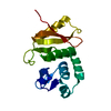
| ||||||||
|---|---|---|---|---|---|---|---|---|---|
| 1 | 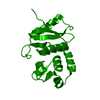
| ||||||||
| Unit cell |
|
-Components
| #1: Protein | Mass: 18448.547 Da / Num. of mol.: 1 Source method: isolated from a genetically manipulated source Source: (gene. exp.) Nanoarchaeum equitans (archaea) / Gene: NEQ261 / Plasmid: pET28a / Production host:  |
|---|
-Experimental details
-Experiment
| Experiment | Method: X-RAY DIFFRACTION / Number of used crystals: 1 |
|---|
- Sample preparation
| Crystal | Density Matthews: 3.41 Å3/Da / Density % sol: 63.88 % |
|---|---|
| Crystal grow | Temperature: 303 K / Method: vapor diffusion, hanging drop / pH: 4.6 Details: 0.10 M sodium acetate pH 4.6, 0.30 M ammonium acetate, 35% PEG 4000, VAPOR DIFFUSION, HANGING DROP, temperature 303K |
-Data collection
| Diffraction | Mean temperature: 100 K |
|---|---|
| Diffraction source | Source: SYNCHROTRON / Site: APS  / Beamline: 22-BM / Wavelength: 1 Å / Beamline: 22-BM / Wavelength: 1 Å |
| Detector | Type: MARMOSAIC 225 mm CCD / Detector: CCD / Date: Mar 14, 2009 |
| Radiation | Monochromator: Si 220 / Protocol: SINGLE WAVELENGTH / Monochromatic (M) / Laue (L): M / Scattering type: x-ray |
| Radiation wavelength | Wavelength: 1 Å / Relative weight: 1 |
| Reflection | Resolution: 2.2→100 Å / Num. all: 13062 / Num. obs: 13062 / % possible obs: 100 % / Observed criterion σ(F): 2 / Observed criterion σ(I): 8 / Redundancy: 19 % / Rmerge(I) obs: 0.097 / Rsym value: 0.097 / Net I/σ(I): 10.5 |
| Reflection shell | Resolution: 2.2→2.28 Å / Redundancy: 11.5 % / Rmerge(I) obs: 0.624 / Mean I/σ(I) obs: 4.9 / Rsym value: 0.624 / % possible all: 99.9 |
-Phasing
| Phasing | Method: molecular replacement |
|---|
- Processing
| Software |
| ||||||||||||||||||||||||||||||||||||||||||||||||||||||||||||||||||||||||||||||||||||||||||||||||||||||||||||||||||||||||||||||||||||||||||||||||||||||||||||||||||||||||||
|---|---|---|---|---|---|---|---|---|---|---|---|---|---|---|---|---|---|---|---|---|---|---|---|---|---|---|---|---|---|---|---|---|---|---|---|---|---|---|---|---|---|---|---|---|---|---|---|---|---|---|---|---|---|---|---|---|---|---|---|---|---|---|---|---|---|---|---|---|---|---|---|---|---|---|---|---|---|---|---|---|---|---|---|---|---|---|---|---|---|---|---|---|---|---|---|---|---|---|---|---|---|---|---|---|---|---|---|---|---|---|---|---|---|---|---|---|---|---|---|---|---|---|---|---|---|---|---|---|---|---|---|---|---|---|---|---|---|---|---|---|---|---|---|---|---|---|---|---|---|---|---|---|---|---|---|---|---|---|---|---|---|---|---|---|---|---|---|---|---|---|---|
| Refinement | Method to determine structure: MOLECULAR REPLACEMENT Starting model: PDB entry 3IEY Resolution: 2.2→41.38 Å / Cor.coef. Fo:Fc: 0.929 / Cor.coef. Fo:Fc free: 0.925 / Occupancy max: 1 / Occupancy min: 1 / SU B: 14.168 / SU ML: 0.162 / TLS residual ADP flag: LIKELY RESIDUAL / Isotropic thermal model: Isotropic / Cross valid method: THROUGHOUT / ESU R: 0.232 / ESU R Free: 0.199 / Stereochemistry target values: MAXIMUM LIKELIHOOD / Details: HYDROGENS HAVE BEEN ADDED IN THE RIDING POSITIONS
| ||||||||||||||||||||||||||||||||||||||||||||||||||||||||||||||||||||||||||||||||||||||||||||||||||||||||||||||||||||||||||||||||||||||||||||||||||||||||||||||||||||||||||
| Solvent computation | Ion probe radii: 0.8 Å / Shrinkage radii: 0.8 Å / VDW probe radii: 1.4 Å / Solvent model: MASK | ||||||||||||||||||||||||||||||||||||||||||||||||||||||||||||||||||||||||||||||||||||||||||||||||||||||||||||||||||||||||||||||||||||||||||||||||||||||||||||||||||||||||||
| Displacement parameters | Biso mean: 35.247 Å2
| ||||||||||||||||||||||||||||||||||||||||||||||||||||||||||||||||||||||||||||||||||||||||||||||||||||||||||||||||||||||||||||||||||||||||||||||||||||||||||||||||||||||||||
| Refinement step | Cycle: LAST / Resolution: 2.2→41.38 Å
| ||||||||||||||||||||||||||||||||||||||||||||||||||||||||||||||||||||||||||||||||||||||||||||||||||||||||||||||||||||||||||||||||||||||||||||||||||||||||||||||||||||||||||
| Refine LS restraints |
| ||||||||||||||||||||||||||||||||||||||||||||||||||||||||||||||||||||||||||||||||||||||||||||||||||||||||||||||||||||||||||||||||||||||||||||||||||||||||||||||||||||||||||
| LS refinement shell | Resolution: 2.2→2.257 Å / Total num. of bins used: 20
| ||||||||||||||||||||||||||||||||||||||||||||||||||||||||||||||||||||||||||||||||||||||||||||||||||||||||||||||||||||||||||||||||||||||||||||||||||||||||||||||||||||||||||
| Refinement TLS params. | Method: refined / Origin x: 27.6367 Å / Origin y: -21.9329 Å / Origin z: 6.3322 Å
|
