 Movie
Movie Controller

[English] 日本語
 Yorodumi
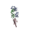
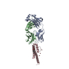
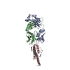
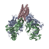
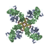
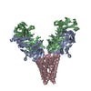
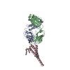
- PDB-2jk5: Potassium Channel KcsA in complex with Tetrabutylammonium in high K -
+ Open data

- Basic information
| Entry | Database: PDB / ID: 2jk5 | ||||||
|---|---|---|---|---|---|---|---|
| Title | Potassium Channel KcsA in complex with Tetrabutylammonium in high K | ||||||
 Components Components |
| ||||||
 Keywords Keywords | IMMUNE SYSTEM/METAL TRANSPORT / IMMUNE SYSTEM-METAL TRANSPORT COMPLEX / QUATERNARY AMMONIUM / PROTEIN-ANTIBODY FAB COMPLEX / IONIC CHANNEL / ION TRANSPORT | ||||||
| Function / homology |  Function and homology information Function and homology informationaction potential / voltage-gated potassium channel activity / voltage-gated potassium channel complex / identical protein binding Similarity search - Function | ||||||
| Biological species |   STREPTOMYCES LIVIDANS (bacteria) STREPTOMYCES LIVIDANS (bacteria) | ||||||
| Method |  X-RAY DIFFRACTION / SYNCHROTRON / MOLECULAR REPLACEMENT / Resolution: 2.4 Å X-RAY DIFFRACTION / SYNCHROTRON / MOLECULAR REPLACEMENT / Resolution: 2.4 Å | ||||||
 Authors Authors | Lenaeus, M.J. / Focia, P.J. / Wagner, T. / Gross, A. | ||||||
 Citation Citation | Journal: Biochemistry / Year: 2014 Title: Structures of Kcsa in Complex with Symmetrical Quaternary Ammonium Compounds Reveal a Hydrophobic Binding Site. Authors: Lenaeus, M.J. / Burdette, D. / Wagner, T. / Focia, P.J. / Gross, A. | ||||||
| History |
| ||||||
| Remark 700 | SHEET THE SHEET STRUCTURE OF THIS MOLECULE IS BIFURCATED. IN ORDER TO REPRESENT THIS FEATURE IN ... SHEET THE SHEET STRUCTURE OF THIS MOLECULE IS BIFURCATED. IN ORDER TO REPRESENT THIS FEATURE IN THE SHEET RECORDS BELOW, TWO SHEETS ARE DEFINED. |
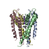
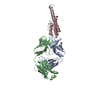
- Structure visualization
| Structure viewer | Molecule: MolmilJmol/JSmol |
|---|
- Downloads & links
-Download
| PDBx/mmCIF format | 2jk5.cif.gz | 126.2 KB | Display | PDBx/mmCIF format |
|---|---|---|---|---|
| PDB format | pdb2jk5.ent.gz | 95.9 KB | Display | PDB format |
| PDBx/mmJSON format | 2jk5.json.gz | Tree view | PDBx/mmJSON format | |
| Others |  Other downloads Other downloads |
-Validation report
| Arichive directory | https://data.pdbj.org/pub/pdb/validation_reports/jk/2jk5ftp://data.pdbj.org/pub/pdb/validation_reports/jk/2jk5 | HTTPS FTP |
|---|
-Related structure data
| Related structure data |  2w0fC 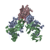 4uujC  1k4cS S: Starting model for refinement C: citing same article ( |
|---|---|
| Similar structure data |


-Links
 PDBj
PDBj


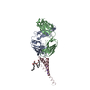
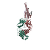
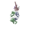
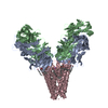
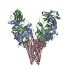
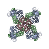
- Assembly
| Deposited unit | 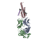
| ||||||||||||||||||||||||||||||
|---|---|---|---|---|---|---|---|---|---|---|---|---|---|---|---|---|---|---|---|---|---|---|---|---|---|---|---|---|---|---|---|
| 1 | 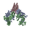
| ||||||||||||||||||||||||||||||
| Unit cell |
| ||||||||||||||||||||||||||||||
| Components on special symmetry positions |
|
-Components
-Protein , 1 types, 1 molecules C
| #3: Protein | Mass: 13237.618 Da / Num. of mol.: 1 / Fragment: RESIDUES 1-124 Source method: isolated from a genetically manipulated source Source: (gene. exp.) STREPTOMYCES LIVIDANS (bacteria) / Production host: |
|---|
-Antibody , 2 types, 2 molecules AB
| #1: Antibody | Mass: 23411.242 Da / Num. of mol.: 1 Source method: isolated from a genetically manipulated source Source: (gene. exp.) |
|---|---|
| #2: Antibody | Mass: 23435.738 Da / Num. of mol.: 1 Source method: isolated from a genetically manipulated source Source: (gene. exp.) |
-Non-polymers , 7 types, 215 molecules 


| #4: Chemical | ChemComp-CO /  Mass: 58.933 Da / Num. of mol.: 1 / Source method: obtained synthetically / Formula: Co Mass: 58.933 Da / Num. of mol.: 1 / Source method: obtained synthetically / Formula: Co | ||||||||||
|---|---|---|---|---|---|---|---|---|---|---|---|
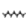
| #5: Chemical | ChemComp-K /  Mass: 39.098 Da / Num. of mol.: 6 / Source method: obtained synthetically / Formula: K Mass: 39.098 Da / Num. of mol.: 6 / Source method: obtained synthetically / Formula: K#6: Chemical | ChemComp-F09 / |  Mass: 144.254 Da / Num. of mol.: 1 / Source method: obtained synthetically / Formula: C9H20O Mass: 144.254 Da / Num. of mol.: 1 / Source method: obtained synthetically / Formula: C9H20O#7: Chemical | ChemComp-L2C / ( |  Mass: 414.619 Da / Num. of mol.: 1 / Source method: obtained synthetically / Formula: C24H46O5 Mass: 414.619 Da / Num. of mol.: 1 / Source method: obtained synthetically / Formula: C24H46O5#8: Chemical | ChemComp-TBA / |  Mass: 242.464 Da / Num. of mol.: 1 / Source method: obtained synthetically / Formula: C16H36N Mass: 242.464 Da / Num. of mol.: 1 / Source method: obtained synthetically / Formula: C16H36N#9: Chemical | ChemComp-HP6 / |  Mass: 100.202 Da / Num. of mol.: 1 / Source method: obtained synthetically / Formula: C7H16 Mass: 100.202 Da / Num. of mol.: 1 / Source method: obtained synthetically / Formula: C7H16#10: Water | ChemComp-HOH / | Mass: 18.015 Da / Num. of mol.: 204 / Source method: isolated from a natural source / Formula: H2O |
-Details
| Has protein modification | Y |
|---|
-Experimental details
-Experiment
| Experiment | Method: X-RAY DIFFRACTION / Number of used crystals: 1 |
|---|
- Sample preparation
| Crystal | Density Matthews: 3.71 Å3/Da / Density % sol: 66.8 % / Description: NONE |
|---|---|
| Crystal grow | pH: 5.4 Details: 18-25%PEG400, 50 MM SODIUM ACETATE, 50 MM MAGNESIUM ACETATE, pH 5.4 |
-Data collection
| Diffraction | Mean temperature: 100 K |
|---|---|
| Diffraction source | Source: SYNCHROTRON / Site: APS  / Beamline: 22-ID / Wavelength: 0.977 / Beamline: 22-ID / Wavelength: 0.977 |
| Detector | Type: MARRESEARCH / Detector: CCD / Date: Nov 19, 2007 |
| Radiation | Protocol: SINGLE WAVELENGTH / Monochromatic (M) / Laue (L): M / Scattering type: x-ray |
| Radiation wavelength | Wavelength: 0.977 Å / Relative weight: 1 |
| Reflection | Resolution: 2.4→50 Å / Num. obs: 35756 / % possible obs: 99.5 % / Observed criterion σ(I): -3 / Redundancy: 3.75 % / Rmerge(I) obs: 0.07 / Net I/σ(I): 15.27 |
| Reflection shell | Resolution: 2.4→2.54 Å / Redundancy: 3.75 % / Rmerge(I) obs: 0.44 / Mean I/σ(I) obs: 3.5 / % possible all: 98.9 |
- Processing
| Software |
| ||||||||||||||||||||||||||||||||||||||||||||||||||||||||||||||||||||||||||||||||||||||||||||||||||||||||||||||||||||||||||||||||||||||||||||||||||||||||||||||||||||||||||||||||||||||
|---|---|---|---|---|---|---|---|---|---|---|---|---|---|---|---|---|---|---|---|---|---|---|---|---|---|---|---|---|---|---|---|---|---|---|---|---|---|---|---|---|---|---|---|---|---|---|---|---|---|---|---|---|---|---|---|---|---|---|---|---|---|---|---|---|---|---|---|---|---|---|---|---|---|---|---|---|---|---|---|---|---|---|---|---|---|---|---|---|---|---|---|---|---|---|---|---|---|---|---|---|---|---|---|---|---|---|---|---|---|---|---|---|---|---|---|---|---|---|---|---|---|---|---|---|---|---|---|---|---|---|---|---|---|---|---|---|---|---|---|---|---|---|---|---|---|---|---|---|---|---|---|---|---|---|---|---|---|---|---|---|---|---|---|---|---|---|---|---|---|---|---|---|---|---|---|---|---|---|---|---|---|---|---|
| Refinement | Method to determine structure: MOLECULAR REPLACEMENT Starting model: PDB ENTRY 1K4C WITH SELECTIVITY FILTER AND IONS REMOVED Resolution: 2.4→30 Å / Cor.coef. Fo:Fc: 0.939 / Cor.coef. Fo:Fc free: 0.908 / SU B: 6.809 / SU ML: 0.158 / Cross valid method: THROUGHOUT / ESU R: 0.259 / ESU R Free: 0.22 / Stereochemistry target values: MAXIMUM LIKELIHOOD Details: SEVERAL DISORDERED REGIONS HAVE BEEN MODELED AS WATER (NEAR TRP B163, CHAIN C22-35, AND THE L2C MOLECULE).
| ||||||||||||||||||||||||||||||||||||||||||||||||||||||||||||||||||||||||||||||||||||||||||||||||||||||||||||||||||||||||||||||||||||||||||||||||||||||||||||||||||||||||||||||||||||||
| Solvent computation | Ion probe radii: 0.8 Å / Shrinkage radii: 0.8 Å / VDW probe radii: 1.2 Å / Solvent model: MASK | ||||||||||||||||||||||||||||||||||||||||||||||||||||||||||||||||||||||||||||||||||||||||||||||||||||||||||||||||||||||||||||||||||||||||||||||||||||||||||||||||||||||||||||||||||||||
| Displacement parameters | Biso mean: 44.2 Å2
| ||||||||||||||||||||||||||||||||||||||||||||||||||||||||||||||||||||||||||||||||||||||||||||||||||||||||||||||||||||||||||||||||||||||||||||||||||||||||||||||||||||||||||||||||||||||
| Refinement step | Cycle: LAST / Resolution: 2.4→30 Å
| ||||||||||||||||||||||||||||||||||||||||||||||||||||||||||||||||||||||||||||||||||||||||||||||||||||||||||||||||||||||||||||||||||||||||||||||||||||||||||||||||||||||||||||||||||||||
| Refine LS restraints |
|