 Movie
Movie Controller


+ Open data

- Basic information
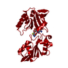
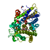
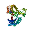
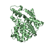
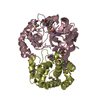
| Entry | Database: PDB / ID: 1bl8 | ||||||
|---|---|---|---|---|---|---|---|
| Title | POTASSIUM CHANNEL (KCSA) FROM STREPTOMYCES LIVIDANS | ||||||
 Components Components | PROTEIN (POTASSIUM CHANNEL PROTEIN) | ||||||
 Keywords Keywords | MEMBRANE PROTEIN / POTASSIUM CHANNEL / INTEGRAL MEMBRANE PROTEIN | ||||||
| Function / homology |  Function and homology information Function and homology informationaction potential / voltage-gated potassium channel activity / voltage-gated potassium channel complex / identical protein binding Similarity search - Function | ||||||
| Biological species |  Streptomyces lividans (bacteria) Streptomyces lividans (bacteria) | ||||||
| Method |  X-RAY DIFFRACTION / SYNCHROTRON / MIR / Resolution: 3.2 Å X-RAY DIFFRACTION / SYNCHROTRON / MIR / Resolution: 3.2 Å | ||||||
 Authors Authors | Doyle, D.A. / Cabral, J.M. / Pfuetzner, R.A. / Kuo, A. / Gulbis, J.M. / Cohen, S.L. / Chait, B.T. / Mackinnon, R. | ||||||
 Citation Citation | Journal: Science / Year: 1998 Title: The structure of the potassium channel: molecular basis of K+ conduction and selectivity. Authors: Doyle, D.A. / Morais Cabral, J. / Pfuetzner, R.A. / Kuo, A. / Gulbis, J.M. / Cohen, S.L. / Chait, B.T. / MacKinnon, R. | ||||||
| History |
|
- Structure visualization
| Structure viewer | Molecule: MolmilJmol/JSmol |
|---|
- Downloads & links
-Download
| PDBx/mmCIF format | 1bl8.cif.gz | 74.9 KB | Display | PDBx/mmCIF format |
|---|---|---|---|---|
| PDB format | pdb1bl8.ent.gz | 58.3 KB | Display | PDB format |
| PDBx/mmJSON format | 1bl8.json.gz | Tree view | PDBx/mmJSON format | |
| Others |  Other downloads Other downloads |
-Validation report
| Arichive directory | https://data.pdbj.org/pub/pdb/validation_reports/bl/1bl8ftp://data.pdbj.org/pub/pdb/validation_reports/bl/1bl8 | HTTPS FTP |
|---|
-Related structure data
| Similar structure data |
|---|


-Links
 PDBj
PDBj

- Assembly
| Deposited unit | 
| ||||||||||||||||
|---|---|---|---|---|---|---|---|---|---|---|---|---|---|---|---|---|---|
| 1 |
| ||||||||||||||||
| Unit cell |
| ||||||||||||||||
| Noncrystallographic symmetry (NCS) | NCS oper:
|
-Components
| #1: Protein | Mass: 10252.959 Da / Num. of mol.: 4 / Mutation: L90C Source method: isolated from a genetically manipulated source Source: (gene. exp.) Streptomyces lividans (bacteria) / Plasmid: PQE60 / Gene (production host): KCSA / Production host: #2: Chemical |   Mass: 39.098 Da / Num. of mol.: 3 / Source method: obtained synthetically / Formula: K Mass: 39.098 Da / Num. of mol.: 3 / Source method: obtained synthetically / Formula: K#3: Water | ChemComp-HOH / |  Mass: 18.015 Da / Num. of mol.: 1 / Source method: isolated from a natural source / Formula: H2O Mass: 18.015 Da / Num. of mol.: 1 / Source method: isolated from a natural source / Formula: H2O |
|---|
-Experimental details
-Experiment
| Experiment | Method: X-RAY DIFFRACTION / Number of used crystals: 6 |
|---|
- Sample preparation
| Crystal | Density Matthews: 4.99 Å3/Da / Density % sol: 75.34 % | ||||||||||||||||||||||||||||||||||||||||||||||||
|---|---|---|---|---|---|---|---|---|---|---|---|---|---|---|---|---|---|---|---|---|---|---|---|---|---|---|---|---|---|---|---|---|---|---|---|---|---|---|---|---|---|---|---|---|---|---|---|---|---|
| Crystal grow | pH: 7.5 Details: ONE-TO-MIXTURE OF PROTEIN SOLUTION AND RESERVOIR (200 MM CACL2, 100 MM HEPES PH 7.5, 48% PEG 400). | ||||||||||||||||||||||||||||||||||||||||||||||||
| Crystal grow | *PLUS Temperature: 20 ℃ / Method: vapor diffusion, sitting dropDetails: drop consists of equal volume of protein and reservoir solutions | ||||||||||||||||||||||||||||||||||||||||||||||||
| Components of the solutions | *PLUS
|
-Data collection
| Diffraction | Mean temperature: 100 K |
|---|---|
| Diffraction source | Source: SYNCHROTRON / Site: CHESS  / Beamline: A1 / Wavelength: 0.908 / Beamline: A1 / Wavelength: 0.908 |
| Detector | Type: PRINCETON 2K / Detector: CCD |
| Radiation | Protocol: SINGLE WAVELENGTH / Monochromatic (M) / Laue (L): M / Scattering type: x-ray |
| Radiation wavelength | Wavelength: 0.908 Å / Relative weight: 1 |
| Reflection | Resolution: 3.2→30 Å / Num. obs: 12603 / % possible obs: 93.3 % / Observed criterion σ(I): 2 / Redundancy: 6.1 % / Biso Wilson estimate: 90 Å2 / Rmerge(I) obs: 0.086 |
| Reflection shell | Resolution: 3.2→3.3 Å / Redundancy: 2.3 % / % possible all: 66.6 |
| Reflection shell | *PLUS % possible obs: 66.6 % / Rmerge(I) obs: 0.286 / Mean I/σ(I) obs: 3.9 |
- Processing
| Software |
| ||||||||||||||||||||||||||||||||||||||||||||||||||||||||||||
|---|---|---|---|---|---|---|---|---|---|---|---|---|---|---|---|---|---|---|---|---|---|---|---|---|---|---|---|---|---|---|---|---|---|---|---|---|---|---|---|---|---|---|---|---|---|---|---|---|---|---|---|---|---|---|---|---|---|---|---|---|---|
| Refinement | Method to determine structure: MIR / Resolution: 3.2→10 Å / Isotropic thermal model: GROUP / σ(F): 2
| ||||||||||||||||||||||||||||||||||||||||||||||||||||||||||||
| Refinement step | Cycle: LAST / Resolution: 3.2→10 Å
| ||||||||||||||||||||||||||||||||||||||||||||||||||||||||||||
| Refine LS restraints |
| ||||||||||||||||||||||||||||||||||||||||||||||||||||||||||||
| Refine LS restraints NCS | NCS model details: RESTRAINTS | ||||||||||||||||||||||||||||||||||||||||||||||||||||||||||||
| Software | *PLUS Name: X-PLOR / Version: 3.851 / Classification: refinement | ||||||||||||||||||||||||||||||||||||||||||||||||||||||||||||
| Refine LS restraints | *PLUS
|
