[English] 日本語
Yorodumi
- PDB-3ifx: Crystal structure of the Spin-labeled KcsA mutant V48R1 -

+
Open data


ID or keywords:

Loading...

-
Basic information

Entry
Database: PDB / ID: 3ifx
TitleCrystal structure of the Spin-labeled KcsA mutant V48R1
ComponentsVoltage-gated potassium channel
KeywordsMEMBRANE PROTEIN / potassium channel / spin-labeled protein / Cell membrane / Ion transport / Ionic channel / Membrane / Transmembrane / Transport / Voltage-gated channel
Function / homology
Function and homology information


monoatomic ion transmembrane transport / identical protein binding / plasma membrane
Similarity search - Function
Helix Hairpins - #70 / Potassium channel domain / Ion channel / Helix Hairpins / Orthogonal Bundle / Mainly Alpha
Similarity search - Domain/homology
: / Chem-MTN / TETRABUTYLAMMONIUM ION / pH-gated potassium channel KcsA
Similarity search - Component
Biological speciesStreptomyces lividans (bacteria)
MethodX-RAY DIFFRACTION / EPR / SYNCHROTRON / MOLECULAR REPLACEMENT / Resolution: 3.56 Å
AuthorsCieslak, J.A. / Focia, P.J. / Gross, A.
CitationJournal: Biochemistry / Year: 2010
Title: Electron Spin-Echo Envelope Modulation (ESEEM) Reveals Water and Phosphate Interactions with the KcsA Potassium Channel
Authors: Cieslak, J.A. / Focia, P.J. / Gross, A.
History
DepositionJul 26, 2009Deposition site: RCSB / Processing site: RCSB
Revision 1.0Feb 9, 2010Provider: repository / Type: Initial release
Revision 1.1Jul 13, 2011Group: Version format compliance
Revision 1.2Jul 27, 2011Group: Advisory / Non-polymer description / Refinement description
Revision 1.3Sep 6, 2023Group: Advisory / Data collection ...Advisory / Data collection / Database references / Derived calculations / Refinement description
Category: chem_comp_atom / chem_comp_bond ...chem_comp_atom / chem_comp_bond / database_2 / pdbx_initial_refinement_model / pdbx_struct_conn_angle / pdbx_unobs_or_zero_occ_atoms / struct_conn / struct_conn_type / struct_ref_seq_dif / struct_site
Item: _database_2.pdbx_DOI / _database_2.pdbx_database_accession ..._database_2.pdbx_DOI / _database_2.pdbx_database_accession / _pdbx_struct_conn_angle.ptnr1_auth_asym_id / _pdbx_struct_conn_angle.ptnr1_auth_comp_id / _pdbx_struct_conn_angle.ptnr1_auth_seq_id / _pdbx_struct_conn_angle.ptnr1_label_asym_id / _pdbx_struct_conn_angle.ptnr1_label_atom_id / _pdbx_struct_conn_angle.ptnr1_label_comp_id / _pdbx_struct_conn_angle.ptnr1_label_seq_id / _pdbx_struct_conn_angle.ptnr2_auth_asym_id / _pdbx_struct_conn_angle.ptnr2_auth_seq_id / _pdbx_struct_conn_angle.ptnr2_label_asym_id / _pdbx_struct_conn_angle.ptnr3_auth_asym_id / _pdbx_struct_conn_angle.ptnr3_auth_comp_id / _pdbx_struct_conn_angle.ptnr3_auth_seq_id / _pdbx_struct_conn_angle.ptnr3_label_asym_id / _pdbx_struct_conn_angle.ptnr3_label_atom_id / _pdbx_struct_conn_angle.ptnr3_label_comp_id / _pdbx_struct_conn_angle.ptnr3_label_seq_id / _pdbx_struct_conn_angle.value / _struct_conn.conn_type_id / _struct_conn.id / _struct_conn.pdbx_dist_value / _struct_conn.pdbx_leaving_atom_flag / _struct_conn.ptnr1_auth_asym_id / _struct_conn.ptnr1_auth_comp_id / _struct_conn.ptnr1_auth_seq_id / _struct_conn.ptnr1_label_asym_id / _struct_conn.ptnr1_label_atom_id / _struct_conn.ptnr1_label_comp_id / _struct_conn.ptnr1_label_seq_id / _struct_conn.ptnr2_auth_asym_id / _struct_conn.ptnr2_auth_comp_id / _struct_conn.ptnr2_auth_seq_id / _struct_conn.ptnr2_label_asym_id / _struct_conn.ptnr2_label_atom_id / _struct_conn.ptnr2_label_comp_id / _struct_conn.ptnr2_label_seq_id / _struct_conn_type.id / _struct_ref_seq_dif.details / _struct_site.pdbx_auth_asym_id / _struct_site.pdbx_auth_comp_id / _struct_site.pdbx_auth_seq_id

-
Structure visualization

Structure viewerMolecule:
MolmilJmol/JSmol

Downloads & links

-
Assembly

Deposited unit
A: Voltage-gated potassium channel
B: Voltage-gated potassium channel
C: Voltage-gated potassium channel
D: Voltage-gated potassium channel
hetero molecules


Theoretical massNumber of molelcules
Total (without water)57,14811
Polymers55,7704
Non-polymers1,3787
Water543
1


  • Idetical with deposited unit
  • defined by author&software
TypeNameSymmetry operationNumber
identity operation1_555x,y,z1
Buried area6930 Å2
ΔGint-71.8 kcal/mol
Surface area17840 Å2
MethodPISA
2
A: Voltage-gated potassium channel
B: Voltage-gated potassium channel
C: Voltage-gated potassium channel
D: Voltage-gated potassium channel
hetero molecules

A: Voltage-gated potassium channel
B: Voltage-gated potassium channel
C: Voltage-gated potassium channel
D: Voltage-gated potassium channel
hetero molecules


Theoretical massNumber of molelcules
Total (without water)114,29522
Polymers111,5398
Non-polymers2,75614
Water724
TypeNameSymmetry operationNumber
identity operation1_555x,y,z1
crystal symmetry operation2_656-x+1,y,-z+11
Buried area15600 Å2
ΔGint-157.6 kcal/mol
Surface area33960 Å2
MethodPISA
Unit cell
Length a, b, c (Å)130.970, 76.630, 112.970
Angle α, β, γ (deg.)90.00, 125.83, 90.00
Int Tables number5
Space group name H-MC121
Details
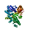
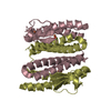
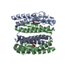
AUTHORS STATE THAT THE QUATERNARY STRUCTURE DEFINED IN REMARK 350 AS BIOMOLECULE 1 IS THE CORRECT PHYSIOLOGICAL TETRAMER THAT FORMS AN ASYMMETRIC UNIT. THE QUATERNARY STRUCTURE DEFINED IN REMARK 350 AS BIOMOLECULE 2 IS INCORRECT AS THE OCTAMERIC STRUCTURE IS A CONSEQUENCE OF CRYSTAL PACKING AND FORMATION OF THE CONTENTS OF THE UNIT CELL.

-
Components

#1: Protein
Voltage-gated potassium channel /


Mass: 13942.380 Da / Num. of mol.: 4 / Fragment: Pore domain: UNP residues 1-124 / Mutation: V48C
Source method: isolated from a genetically manipulated source
Source: (gene. exp.) Streptomyces lividans (bacteria) / Gene: kcsA, skc1 / Plasmid: pQE60 / Production host: Escherichia coli (E. coli) / References: UniProt: P0A334
#2: Chemical ChemComp-K / POTASSIUM ION


Mass: 39.098 Da / Num. of mol.: 2 / Source method: obtained synthetically / Formula: K
#3: Chemical
ChemComp-MTN / S-[(1-oxyl-2,2,5,5-tetramethyl-2,5-dihydro-1H-pyrrol-3-yl)methyl] methanesulfonothioate / MTSL / MTSL


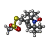
Mass: 264.385 Da / Num. of mol.: 4 / Source method: obtained synthetically / Formula: C10H18NO3S2
#4: Chemical ChemComp-TBA / TETRABUTYLAMMONIUM ION / Tetrabutylammonium


Mass: 242.464 Da / Num. of mol.: 1 / Source method: obtained synthetically / Formula: C16H36N
#5: Water ChemComp-HOH / water / Water


Mass: 18.015 Da / Num. of mol.: 3 / Source method: isolated from a natural source / Formula: H2O

-
Experimental details

-
Experiment

Experiment
MethodNumber of used crystals
X-RAY DIFFRACTION1
EPR1

-
Sample preparation

CrystalDensity Matthews: 4.28 Å3/Da / Density % sol: 71.23 %
Crystal growTemperature: 293 K / Method: vapor diffusion, sitting drop / pH: 7.5
Details: 200 mM CaCl2, 150 mM KCl, 100 mM HEPES, 19-49% PEG 400, pH 7.5, VAPOR DIFFUSION, SITTING DROP, temperature 293K

-
Data collection

DiffractionMean temperature: 100 K
Diffraction sourceSource: SYNCHROTRON / Site: APS / Beamline: 23-ID-B / Wavelength: 1.033 Å
DetectorType: MARMOSAIC 300 mm CCD / Detector: CCD / Date: Dec 15, 2008
Details: K-B pair of biomorph mirrors for vertical and horizontal focusing
RadiationMonochromator: Si(111) double crystal / Protocol: SINGLE WAVELENGTH / Monochromatic (M) / Laue (L): M / Scattering type: x-ray
Radiation wavelengthWavelength: 1.033 Å / Relative weight: 1
ReflectionResolution: 3.56→50 Å / Num. all: 7946 / Num. obs: 7898 / % possible obs: 99.4 % / Observed criterion σ(F): 1 / Redundancy: 3.5 % / Rmerge(I) obs: 0.086 / Net I/σ(I): 4
Reflection shellResolution: 3.56→3.65 Å / Redundancy: 3.6 % / Rmerge(I) obs: 0.0421 / Mean I/σ(I) obs: 1.7 / % possible all: 99.4

-
Processing

Software
NameVersionClassification
MOSFLMdata reduction
PHASERphasing
REFMAC5.5.0051refinement
SCALAdata scaling
RefinementMethod to determine structure: MOLECULAR REPLACEMENT
Starting model: PDB entry 1BL8
Resolution: 3.56→24.84 Å / Cor.coef. Fo:Fc: 0.904 / Cor.coef. Fo:Fc free: 0.897 / SU B: 66.443 / SU ML: 0.512 / TLS residual ADP flag: LIKELY RESIDUAL / Cross valid method: THROUGHOUT / σ(F): 0 / ESU R Free: 0.721 / Stereochemistry target values: MAXIMUM LIKELIHOOD / Details: HYDROGENS HAVE BEEN ADDED IN THE RIDING POSITIONS
RfactorNum. reflection% reflectionSelection details
Rfree0.30218 409 4.9 %RANDOM
Rwork0.27109 ---
all0.27267 7898 --
obs0.27267 7898 75.14 %-
Solvent computationIon probe radii: 0.8 Å / Shrinkage radii: 0.8 Å / VDW probe radii: 1.4 Å / Solvent model: MASK
Displacement parametersBiso mean: 110.671 Å2
Baniso -1Baniso -2Baniso -3
1--0.22 Å20 Å2-0.67 Å2
2---0.12 Å20 Å2
3----0.45 Å2
Refinement stepCycle: LAST / Resolution: 3.56→24.84 Å
ProteinNucleic acidLigandSolventTotal
Num. atoms2743 0 49 3 2795
Refine LS restraints
Refine-IDTypeDev idealDev ideal targetNumber
X-RAY DIFFRACTIONr_bond_refined_d0.0550.0222914
X-RAY DIFFRACTIONr_angle_refined_deg1.7791.9644018
X-RAY DIFFRACTIONr_dihedral_angle_1_deg6.335388
X-RAY DIFFRACTIONr_dihedral_angle_2_deg38.9721.85770
X-RAY DIFFRACTIONr_dihedral_angle_3_deg26.17715353
X-RAY DIFFRACTIONr_dihedral_angle_4_deg15.683157
X-RAY DIFFRACTIONr_chiral_restr0.0860.2512
X-RAY DIFFRACTIONr_gen_planes_refined0.0070.0212074
X-RAY DIFFRACTIONr_mcbond_it0.8181.5107
X-RAY DIFFRACTIONr_mcangle_it1.5112165
X-RAY DIFFRACTIONr_scbond_it0.844341
X-RAY DIFFRACTIONr_scangle_it1.6814.540
LS refinement shellResolution: 3.56→3.65 Å / Total num. of bins used: 20
RfactorNum. reflection% reflection
Rfree0.462 5 -
Rwork0.354 66 -
obs--8.78 %
Refinement TLS params.

L11: 0 °2 / L12: 0 °2 / L13: 0 °2 / L22: 0 °2 / L23: 0 °2 / L33: 0 °2 / S11: 0 Å ° / S12: 0 Å ° / S13: 0 Å ° / S21: 0 Å ° / S22: 0 Å ° / S23: 0 Å ° / S31: 0 Å ° / S32: 0 Å ° / S33: 0 Å ° / T11: 0 Å2 / T12: 0 Å2 / T13: 0 Å2 / T22: 0 Å2 / T23: 0 Å2 / T33: 0 Å2 / Method: refined / Refine-ID: X-RAY DIFFRACTION

IDOrigin x (Å)Origin y (Å)Origin z (Å)
136.532929.931921.6098
226.096716.081425.3794
313.41326.824630.2369
423.451940.586126.4115
Refinement TLS group
IDRefine-IDRefine TLS-IDAuth asym-IDAuth seq-ID
1X-RAY DIFFRACTION1B23 - 34
2X-RAY DIFFRACTION1B35 - 56
3X-RAY DIFFRACTION1B57 - 67
4X-RAY DIFFRACTION1B68 - 78
5X-RAY DIFFRACTION1B79 - 89
6X-RAY DIFFRACTION1B90 - 112
7X-RAY DIFFRACTION1B113 - 119
8X-RAY DIFFRACTION2C23 - 36
9X-RAY DIFFRACTION2C37 - 47
10X-RAY DIFFRACTION2C48 - 60
11X-RAY DIFFRACTION2C61 - 65
12X-RAY DIFFRACTION2C66 - 82
13X-RAY DIFFRACTION2C83 - 92
14X-RAY DIFFRACTION2C93 - 110
15X-RAY DIFFRACTION2C111 - 119
16X-RAY DIFFRACTION3D23 - 34
17X-RAY DIFFRACTION3D35 - 56
18X-RAY DIFFRACTION3D57 - 67
19X-RAY DIFFRACTION3D68 - 78
20X-RAY DIFFRACTION3D79 - 89
21X-RAY DIFFRACTION3D90 - 112
22X-RAY DIFFRACTION3D113 - 119
23X-RAY DIFFRACTION4A23 - 36
24X-RAY DIFFRACTION4A37 - 47
25X-RAY DIFFRACTION4A48 - 60
26X-RAY DIFFRACTION4A61 - 65
27X-RAY DIFFRACTION4A66 - 82
28X-RAY DIFFRACTION4A83 - 92
29X-RAY DIFFRACTION4A93 - 110
30X-RAY DIFFRACTION4A111 - 119

+
About Yorodumi

-
News

-
Feb 9, 2022. New format data for meta-information of EMDB entries

New format data for meta-information of EMDB entries

  • Version 3 of the EMDB header file is now the official format.
  • The previous official version 1.9 will be removed from the archive.

Related info.:EMDB header

External links:wwPDB to switch to version 3 of the EMDB data model

-
Aug 12, 2020. Covid-19 info

Covid-19 info

URL: https://pdbj.org/emnavi/covid19.php

New page: Covid-19 featured information page in EM Navigator.

Related info.:Covid-19 info / Mar 5, 2020. Novel coronavirus structure data

+
Mar 5, 2020. Novel coronavirus structure data

Novel coronavirus structure data

Related info.:Yorodumi Speices / Aug 12, 2020. Covid-19 info

External links:COVID-19 featured content - PDBj / Molecule of the Month (242):Coronavirus Proteases

+
Jan 31, 2019. EMDB accession codes are about to change! (news from PDBe EMDB page)

EMDB accession codes are about to change! (news from PDBe EMDB page)

  • The allocation of 4 digits for EMDB accession codes will soon come to an end. Whilst these codes will remain in use, new EMDB accession codes will include an additional digit and will expand incrementally as the available range of codes is exhausted. The current 4-digit format prefixed with “EMD-” (i.e. EMD-XXXX) will advance to a 5-digit format (i.e. EMD-XXXXX), and so on. It is currently estimated that the 4-digit codes will be depleted around Spring 2019, at which point the 5-digit format will come into force.
  • The EM Navigator/Yorodumi systems omit the EMD- prefix.

Related info.:Q: What is EMD? / ID/Accession-code notation in Yorodumi/EM Navigator

External links:EMDB Accession Codes are Changing Soon! / Contact to PDBj

+
Jul 12, 2017. Major update of PDB

Major update of PDB

  • wwPDB released updated PDB data conforming to the new PDBx/mmCIF dictionary.
  • This is a major update changing the version number from 4 to 5, and with Remediation, in which all the entries are updated.
  • In this update, many items about electron microscopy experimental information are reorganized (e.g. em_software).
  • Now, EM Navigator and Yorodumi are based on the updated data.

External links:wwPDB Remediation / Enriched Model Files Conforming to OneDep Data Standards Now Available in the PDB FTP Archive

-
Yorodumi

Thousand views of thousand structures

  • Yorodumi is a browser for structure data from EMDB, PDB, SASBDB, etc.
  • This page is also the successor to EM Navigator detail page, and also detail information page/front-end page for Omokage search.
  • The word "yorodu" (or yorozu) is an old Japanese word meaning "ten thousand". "mi" (miru) is to see.

Related info.:EMDB / PDB / SASBDB / Comparison of 3 databanks / Yorodumi Search / Aug 31, 2016. New EM Navigator & Yorodumi / Yorodumi Papers / Jmol/JSmol / Function and homology information / Changes in new EM Navigator and Yorodumi

Read more