 Movie
Movie Controller


+ Open data

- Basic information
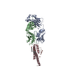
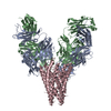
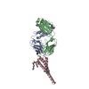
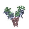
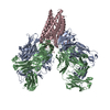
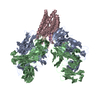
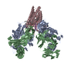
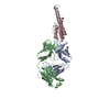
| Entry | Database: PDB / ID: 1r3i | ||||||
|---|---|---|---|---|---|---|---|
| Title | potassium channel KcsA-Fab complex in Rb+ | ||||||
 Components Components |
| ||||||
 Keywords Keywords | MEMBRANE PROTEIN / potassium channel / KcsA-Fab complex / rubidium | ||||||
| Function / homology |  Function and homology information Function and homology informationalpha-beta T cell receptor complex / IgG immunoglobulin complex / action potential / voltage-gated potassium channel activity / voltage-gated potassium channel complex / B cell differentiation / adaptive immune response / extracellular region / identical protein binding / plasma membrane Similarity search - Function | ||||||
| Biological species |  Streptomyces lividans (bacteria) Streptomyces lividans (bacteria) | ||||||
| Method |  X-RAY DIFFRACTION / SYNCHROTRON / MOLECULAR REPLACEMENT / Resolution: 2.4 Å X-RAY DIFFRACTION / SYNCHROTRON / MOLECULAR REPLACEMENT / Resolution: 2.4 Å | ||||||
 Authors Authors | Zhou, Y. / MacKinnon, R. | ||||||
 Citation Citation | Journal: J.Mol.Biol. / Year: 2003 Title: The occupancy of ions in the K+ selectivity filter: Charge balance and coupling of ion binding to a protein conformational change underlie high conduction rates Authors: Zhou, Y. / MacKinnon, R. | ||||||
| History |
|
- Structure visualization
| Structure viewer | Molecule: MolmilJmol/JSmol |
|---|
- Downloads & links
-Download
| PDBx/mmCIF format | 1r3i.cif.gz | 122.8 KB | Display | PDBx/mmCIF format |
|---|---|---|---|---|
| PDB format | pdb1r3i.ent.gz | 91.9 KB | Display | PDB format |
| PDBx/mmJSON format | 1r3i.json.gz | Tree view | PDBx/mmJSON format | |
| Others |  Other downloads Other downloads |
-Validation report
| Arichive directory | https://data.pdbj.org/pub/pdb/validation_reports/r3/1r3iftp://data.pdbj.org/pub/pdb/validation_reports/r3/1r3i | HTTPS FTP |
|---|
-Related structure data
| Related structure data |  1r3jC  1r3kC  1r3lC  1k4cS S: Starting model for refinement C: citing same article ( |
|---|---|
| Similar structure data |


-Links
 PDBj
PDBj


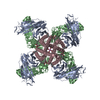
- Assembly
| Deposited unit | 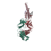
| |||||||||||||||
|---|---|---|---|---|---|---|---|---|---|---|---|---|---|---|---|---|
| 1 | 
| |||||||||||||||
| Unit cell |
| |||||||||||||||
| Components on special symmetry positions |
| |||||||||||||||
| Details | homo-tetramer of KcsA is generated by four fold axis: x,y,z -x,-y,z -x,y,z x,-y,z |
-Components
-Protein , 1 types, 1 molecules C
| #3: Protein | Mass: 13211.582 Da / Num. of mol.: 1 / Mutation: P2A,L90C Source method: isolated from a genetically manipulated source Source: (gene. exp.) Streptomyces lividans (bacteria) / Gene: KCSA, SKC1, SCO7660, SC10F4.33 / Plasmid: pQE60 / Production host: |
|---|
-Antibody , 2 types, 2 molecules LH
| #1: Antibody | Mass: 23435.738 Da / Num. of mol.: 1 / Source method: isolated from a natural source / Source: (natural) |
|---|---|
| #2: Antibody | Mass: 23411.242 Da / Num. of mol.: 1 / Source method: isolated from a natural source / Source: (natural) |
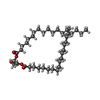
-Non-polymers , 4 types, 178 molecules 


| #4: Chemical | ChemComp-F09 /  Mass: 144.254 Da / Num. of mol.: 1 / Source method: obtained synthetically / Formula: C9H20O Mass: 144.254 Da / Num. of mol.: 1 / Source method: obtained synthetically / Formula: C9H20O | ||||
|---|---|---|---|---|---|
| #5: Chemical | ChemComp-RB /  Mass: 85.468 Da / Num. of mol.: 4 / Source method: obtained synthetically / Formula: Rb Mass: 85.468 Da / Num. of mol.: 4 / Source method: obtained synthetically / Formula: Rb#6: Chemical | ChemComp-DGA / |  Mass: 625.018 Da / Num. of mol.: 1 / Source method: obtained synthetically / Formula: C39H76O5 Mass: 625.018 Da / Num. of mol.: 1 / Source method: obtained synthetically / Formula: C39H76O5#7: Water | ChemComp-HOH / | Mass: 18.015 Da / Num. of mol.: 172 / Source method: isolated from a natural source / Formula: H2O |
-Details
| Has protein modification | Y |
|---|
-Experimental details
-Experiment
| Experiment | Method: X-RAY DIFFRACTION / Number of used crystals: 1 |
|---|
- Sample preparation
| Crystal | Density Matthews: 3.83 Å3/Da / Density % sol: 67.91 % | |||||||||||||||||||||||||||||||||||||||||||||||||
|---|---|---|---|---|---|---|---|---|---|---|---|---|---|---|---|---|---|---|---|---|---|---|---|---|---|---|---|---|---|---|---|---|---|---|---|---|---|---|---|---|---|---|---|---|---|---|---|---|---|---|
| Crystal grow | Temperature: 293 K / Method: vapor diffusion, sitting drop / pH: 5.4 Details: PEG400, sodium acetate, magnesium acetate, pH 5.4, VAPOR DIFFUSION, SITTING DROP, temperature 293K | |||||||||||||||||||||||||||||||||||||||||||||||||
| Crystal grow | *PLUS Temperature: 20 ℃ / pH: 7.5 / Method: vapor diffusion, sitting drop | |||||||||||||||||||||||||||||||||||||||||||||||||
| Components of the solutions | *PLUS
|
-Data collection
| Diffraction | Mean temperature: 100 K |
|---|---|
| Diffraction source | Source: SYNCHROTRON / Site: NSLS  / Beamline: X25 / Wavelength: 1.1 Å / Beamline: X25 / Wavelength: 1.1 Å |
| Detector | Type: BRANDEIS - B4 / Detector: CCD / Date: Jun 17, 2001 |
| Radiation | Monochromator: Si 111 / Protocol: SINGLE WAVELENGTH / Monochromatic (M) / Laue (L): M / Scattering type: x-ray |
| Radiation wavelength | Wavelength: 1.1 Å / Relative weight: 1 |
| Reflection | Resolution: 2.4→30 Å / Num. all: 33943 / Num. obs: 33943 / % possible obs: 99.1 % / Redundancy: 3.4 % / Biso Wilson estimate: 42.9 Å2 / Rmerge(I) obs: 0.074 / Net I/σ(I): 15.2 |
| Reflection shell | Resolution: 2.4→2.5 Å / Redundancy: 2.5 % / Rmerge(I) obs: 0.326 / Mean I/σ(I) obs: 2.6 / Num. unique all: 3185 / % possible all: 93.7 |
- Processing
| Software |
| |||||||||||||||||||||||||
|---|---|---|---|---|---|---|---|---|---|---|---|---|---|---|---|---|---|---|---|---|---|---|---|---|---|---|
| Refinement | Method to determine structure: MOLECULAR REPLACEMENT Starting model: 1K4C Resolution: 2.4→30 Å / Cross valid method: THROUGHOUT / σ(F): 0 / Stereochemistry target values: Engh & Huber Details: The occupancy of ions in this model were set to 1. Please refer to the primary citation for a detailed analysis of ion occupancy.
| |||||||||||||||||||||||||
| Refine analyze |
| |||||||||||||||||||||||||
| Refinement step | Cycle: LAST / Resolution: 2.4→30 Å
| |||||||||||||||||||||||||
| Refine LS restraints |
| |||||||||||||||||||||||||
| LS refinement shell | Resolution: 2.43→2.55 Å / Rfactor Rfree error: 0.006
| |||||||||||||||||||||||||
| Refinement | *PLUS Rfactor Rfree: 0.238 / Rfactor Rwork: 0.218 | |||||||||||||||||||||||||
| Solvent computation | *PLUS | |||||||||||||||||||||||||
| Displacement parameters | *PLUS | |||||||||||||||||||||||||
| Refine LS restraints | *PLUS
|
