 Movie
Movie Controller

[English] 日本語
 Yorodumi
- PDB-2jfv: Crystal structure of Enterococcus faecium glutamate racemase in c... -
+ Open data

- Basic information
| Entry | Database: PDB / ID: 2jfv | ||||||
|---|---|---|---|---|---|---|---|
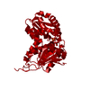
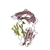
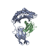
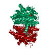
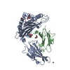
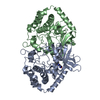
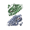
| Title | Crystal structure of Enterococcus faecium glutamate racemase in complex with citrate | ||||||
 Components Components | GLUTAMATE RACEMASE | ||||||
 Keywords Keywords | ISOMERASE / GLUTAMATE RACEMASE / PEPTIDOGLYCAN BIOSYNTHESIS | ||||||
| Function / homology |  Function and homology information Function and homology informationglutamate racemase / glutamate racemase activity / peptidoglycan biosynthetic process / cell wall organization / regulation of cell shape / identical protein binding Similarity search - Function | ||||||
| Biological species |  ENTEROCOCCUS FAECIUM (bacteria) ENTEROCOCCUS FAECIUM (bacteria) | ||||||
| Method |  X-RAY DIFFRACTION / SYNCHROTRON / MOLECULAR REPLACEMENT / Resolution: 1.8 Å X-RAY DIFFRACTION / SYNCHROTRON / MOLECULAR REPLACEMENT / Resolution: 1.8 Å | ||||||
 Authors Authors | Lundqvist, T. | ||||||
 Citation Citation | Journal: Nature / Year: 2007 Title: Exploitation of Structural and Regulatory Diversity in Glutamate Racemases Authors: Lundqvist, T. / Fisher, S.L. / Kern, G. / Folmer, R.H.A. / Xue, Y. / Newton, D.T. / Keating, T.A. / Alm, R.A. / De Jonge, B.L.M. | ||||||
| History |
|
- Structure visualization
| Structure viewer | Molecule: MolmilJmol/JSmol |
|---|
- Downloads & links
-Download
| PDBx/mmCIF format | 2jfv.cif.gz | 71.9 KB | Display | PDBx/mmCIF format |
|---|---|---|---|---|
| PDB format | pdb2jfv.ent.gz | 52 KB | Display | PDB format |
| PDBx/mmJSON format | 2jfv.json.gz | Tree view | PDBx/mmJSON format | |
| Others |  Other downloads Other downloads |
-Validation report
| Arichive directory | https://data.pdbj.org/pub/pdb/validation_reports/jf/2jfvftp://data.pdbj.org/pub/pdb/validation_reports/jf/2jfv | HTTPS FTP |
|---|
-Related structure data
| Related structure data |  2jfnC  2jfoC  2jfpC 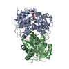 2jfqC  2jfuSC 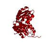 2jfwC  2jfxC  2jfyC 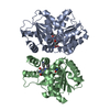 2jfzC S: Starting model for refinement C: citing same article ( |
|---|---|
| Similar structure data |


-Links
 PDBj
PDBj


- Assembly
| Deposited unit | 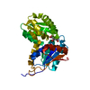
| ||||||||
|---|---|---|---|---|---|---|---|---|---|
| 1 | 
| ||||||||
| Unit cell |
|
-Components
| #1: Protein | Mass: 31664.420 Da / Num. of mol.: 1 / Fragment: RESIDUES 4-277 Source method: isolated from a genetically manipulated source Source: (gene. exp.) ENTEROCOCCUS FAECIUM (bacteria) / Production host: |
|---|---|
| #2: Chemical | ChemComp-FLC /   Mass: 189.100 Da / Num. of mol.: 1 / Source method: obtained synthetically / Formula: C6H5O7 Mass: 189.100 Da / Num. of mol.: 1 / Source method: obtained synthetically / Formula: C6H5O7 |
| #3: Water | ChemComp-HOH /  Mass: 18.015 Da / Num. of mol.: 282 / Source method: isolated from a natural source / Formula: H2O Mass: 18.015 Da / Num. of mol.: 282 / Source method: isolated from a natural source / Formula: H2O |
-Experimental details
-Experiment
| Experiment | Method: X-RAY DIFFRACTION / Number of used crystals: 1 |
|---|
- Sample preparation
| Crystal | Density Matthews: 3.3 Å3/Da / Density % sol: 63 % |
|---|
-Data collection
| Diffraction | Mean temperature: 100 K |
|---|---|
| Diffraction source | Source: SYNCHROTRON / Site: ESRF  / Beamline: ID29 / Wavelength: 0.9793 / Beamline: ID29 / Wavelength: 0.9793 |
| Detector | Type: ADSC CCD / Detector: CCD / Date: May 11, 2003 |
| Radiation | Protocol: SINGLE WAVELENGTH / Monochromatic (M) / Laue (L): M / Scattering type: x-ray |
| Radiation wavelength | Wavelength: 0.9793 Å / Relative weight: 1 |
| Reflection | Resolution: 1.8→34 Å / Num. obs: 34849 / % possible obs: 95.5 % / Observed criterion σ(I): 2 / Redundancy: 4.4 % / Rmerge(I) obs: 0.05 / Net I/σ(I): 10.1 |
| Reflection shell | Resolution: 1.8→1.9 Å / Redundancy: 2 % / Rmerge(I) obs: 0.2 / Mean I/σ(I) obs: 3.7 / % possible all: 95.5 |
- Processing
| Software |
| ||||||||||||||||||||||||||||||||||||||||||||||||||||||||||||
|---|---|---|---|---|---|---|---|---|---|---|---|---|---|---|---|---|---|---|---|---|---|---|---|---|---|---|---|---|---|---|---|---|---|---|---|---|---|---|---|---|---|---|---|---|---|---|---|---|---|---|---|---|---|---|---|---|---|---|---|---|---|
| Refinement | Method to determine structure: MOLECULAR REPLACEMENT Starting model: PDB ENTRY 2JFU Resolution: 1.8→34 Å / Cross valid method: THROUGHOUT / σ(F): 0
| ||||||||||||||||||||||||||||||||||||||||||||||||||||||||||||
| Solvent computation | Bsol: 63.043 Å2 / ksol: 0.445138 e/Å3 | ||||||||||||||||||||||||||||||||||||||||||||||||||||||||||||
| Displacement parameters | Biso mean: 26 Å2
| ||||||||||||||||||||||||||||||||||||||||||||||||||||||||||||
| Refinement step | Cycle: LAST / Resolution: 1.8→34 Å
| ||||||||||||||||||||||||||||||||||||||||||||||||||||||||||||
| Refine LS restraints |
|
