 Movie
Movie Controller

[English] 日本語
 Yorodumi
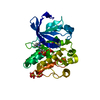
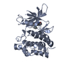
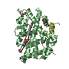
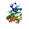
- PDB-2jfn: Crystal structure of Escherichia coli glutamate racemase in compl... -
+ Open data

- Basic information
| Entry | Database: PDB / ID: 2jfn | ||||||
|---|---|---|---|---|---|---|---|
| Title | Crystal structure of Escherichia coli glutamate racemase in complex with L- Glutamate and activator UDP-MurNAc-ala | ||||||
 Components Components | GLUTAMATE RACEMASE | ||||||
 Keywords Keywords | ISOMERASE / CELL WALL / CELL SHAPE / UDP- MURNAC-ALA / PEPTIDOGLYCAN BIOSYNTHESIS / GLUTAMATE RACEMASE / PEPTIDOGLYCAN SYNTHESIS | ||||||
| Function / homology |  Function and homology information Function and homology informationglutamate racemase / glutamate racemase activity / peptidoglycan biosynthetic process / cell wall organization / regulation of cell shape Similarity search - Function | ||||||
| Biological species |  | ||||||
| Method |  X-RAY DIFFRACTION / SYNCHROTRON / MAD / Resolution: 1.9 Å X-RAY DIFFRACTION / SYNCHROTRON / MAD / Resolution: 1.9 Å | ||||||
 Authors Authors | Lundqvist, T. | ||||||
 Citation Citation | Journal: Nature / Year: 2007 Title: Exploitation of Structural and Regulatory Diversity in Glutamate Racemases Authors: Lundqvist, T. / Fisher, S.L. / Kern, G. / Folmer, R.H.A. / Xue, Y. / Newton, D.T. / Keating, T.A. / Alm, R.A. / De Jonge, B.L.M. | ||||||
| History |
| ||||||
| Remark 700 | SHEET THE SHEET STRUCTURE OF THIS MOLECULE IS BIFURCATED. IN ORDER TO REPRESENT THIS FEATURE IN ... SHEET THE SHEET STRUCTURE OF THIS MOLECULE IS BIFURCATED. IN ORDER TO REPRESENT THIS FEATURE IN THE SHEET RECORDS BELOW, TWO SHEETS ARE DEFINED. |
- Structure visualization
| Structure viewer | Molecule: MolmilJmol/JSmol |
|---|
- Downloads & links
-Download
| PDBx/mmCIF format | 2jfn.cif.gz | 69.4 KB | Display | PDBx/mmCIF format |
|---|---|---|---|---|
| PDB format | pdb2jfn.ent.gz | 51.2 KB | Display | PDB format |
| PDBx/mmJSON format | 2jfn.json.gz | Tree view | PDBx/mmJSON format | |
| Others |  Other downloads Other downloads |
-Validation report
| Arichive directory | https://data.pdbj.org/pub/pdb/validation_reports/jf/2jfnftp://data.pdbj.org/pub/pdb/validation_reports/jf/2jfn | HTTPS FTP |
|---|
-Related structure data
| Related structure data |  2jfoC  2jfpC 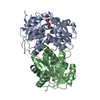 2jfqC 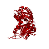 2jfuC 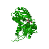 2jfvC 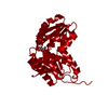 2jfwC  2jfxC  2jfyC 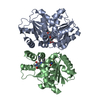 2jfzC C: citing same article ( |
|---|---|
| Similar structure data |


-Links
 PDBj
PDBj

- Assembly
| Deposited unit | 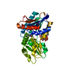
| ||||||||
|---|---|---|---|---|---|---|---|---|---|
| 1 |
| ||||||||
| Unit cell |
|
-Components
| #1: Protein | Mass: 31033.871 Da / Num. of mol.: 1 Source method: isolated from a genetically manipulated source Source: (gene. exp.) |
|---|---|
| #2: Chemical | ChemComp-GLU /   Type: L-peptide linking / Mass: 147.129 Da / Num. of mol.: 1 / Source method: obtained synthetically / Formula: C5H9NO4 Type: L-peptide linking / Mass: 147.129 Da / Num. of mol.: 1 / Source method: obtained synthetically / Formula: C5H9NO4 |
| #3: Chemical | ChemComp-UMA / 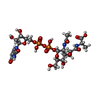  Type: L-peptide linking / Mass: 750.494 Da / Num. of mol.: 1 / Source method: obtained synthetically / Formula: C23H36N4O20P2 Type: L-peptide linking / Mass: 750.494 Da / Num. of mol.: 1 / Source method: obtained synthetically / Formula: C23H36N4O20P2 |
| #4: Water | ChemComp-HOH /  Mass: 18.015 Da / Num. of mol.: 221 / Source method: isolated from a natural source / Formula: H2O Mass: 18.015 Da / Num. of mol.: 221 / Source method: isolated from a natural source / Formula: H2O |
-Experimental details
-Experiment
| Experiment | Method: X-RAY DIFFRACTION / Number of used crystals: 1 |
|---|
- Sample preparation
| Crystal | Density Matthews: 2.66 Å3/Da / Density % sol: 53 % |
|---|---|
| Crystal grow | Details: PROTEIN FORMULATED AT 10 MG/ML WITH 200MM AMMONIUM ACETATE PH 7.4, 5MM D-L GLUTAMATE, 1 MM TCEP AND IN 0.6 MM OF THE ACTIVATOR MUR-NAC-ALA AND CRYSTALLISED WITH 100 MM SODIUM ACETATE PH 4.55- ...Details: PROTEIN FORMULATED AT 10 MG/ML WITH 200MM AMMONIUM ACETATE PH 7.4, 5MM D-L GLUTAMATE, 1 MM TCEP AND IN 0.6 MM OF THE ACTIVATOR MUR-NAC-ALA AND CRYSTALLISED WITH 100 MM SODIUM ACETATE PH 4.55-10% MME 2000 AND 30% GLYCEROL |
-Data collection
| Diffraction | Mean temperature: 100 K |
|---|---|
| Diffraction source | Source: SYNCHROTRON / Site: ESRF  / Beamline: ID14-4 / Wavelength: 0.9456 / Beamline: ID14-4 / Wavelength: 0.9456 |
| Detector | Type: ADSC CCD / Detector: CCD / Date: Apr 11, 2000 |
| Radiation | Protocol: SINGLE WAVELENGTH / Monochromatic (M) / Laue (L): M / Scattering type: x-ray |
| Radiation wavelength | Wavelength: 0.9456 Å / Relative weight: 1 |
| Reflection | Resolution: 1.9→20 Å / Num. obs: 338825 / % possible obs: 95.6 % / Observed criterion σ(I): 2.1 / Redundancy: 6.4 % / Rmerge(I) obs: 0.07 / Net I/σ(I): 5.6 |
| Reflection shell | Resolution: 1.9→2 Å / Redundancy: 3.7 % / Rmerge(I) obs: 0.36 / Mean I/σ(I) obs: 2.1 / % possible all: 78.5 |
- Processing
| Software |
| ||||||||||||||||||||||||||||||||||||||||||||||||||||||||||||
|---|---|---|---|---|---|---|---|---|---|---|---|---|---|---|---|---|---|---|---|---|---|---|---|---|---|---|---|---|---|---|---|---|---|---|---|---|---|---|---|---|---|---|---|---|---|---|---|---|---|---|---|---|---|---|---|---|---|---|---|---|---|
| Refinement | Method to determine structure: MAD / Resolution: 1.9→20 Å / Cross valid method: THROUGHOUT / σ(F): 0
| ||||||||||||||||||||||||||||||||||||||||||||||||||||||||||||
| Solvent computation | Bsol: 48.6342 Å2 / ksol: 0.373583 e/Å3 | ||||||||||||||||||||||||||||||||||||||||||||||||||||||||||||
| Displacement parameters |
| ||||||||||||||||||||||||||||||||||||||||||||||||||||||||||||
| Refinement step | Cycle: LAST / Resolution: 1.9→20 Å
| ||||||||||||||||||||||||||||||||||||||||||||||||||||||||||||
| Refine LS restraints |
| ||||||||||||||||||||||||||||||||||||||||||||||||||||||||||||
| Xplor file | Serial no: 1 / Param file: A / Topol file: A |
