 Movie
Movie Controller

[English] 日本語
 Yorodumi
- PDB-2jf3: Nucleotide substrate binding by UDP-N-acetylglucosamine acyltrans... -
+ Open data

- Basic information
| Entry | Database: PDB / ID: 2jf3 | ||||||
|---|---|---|---|---|---|---|---|
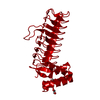
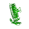
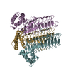
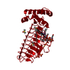
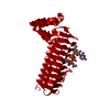
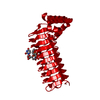
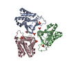
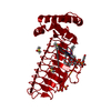
| Title | Nucleotide substrate binding by UDP-N-acetylglucosamine acyltransferase | ||||||
 Components Components | ACYL-[ACYL-CARRIER-PROTEIN]--UDP-N-ACETYLGLUCOSAMINE O-ACYLTRANSFERASE | ||||||
 Keywords Keywords | TRANSFERASE / LIPID A BIOSYNTHESIS / ACYLTRANSFERASE / LIPID SYNTHESIS | ||||||
| Function / homology |  Function and homology information Function and homology informationacyl-[acyl-carrier-protein]-UDP-N-acetylglucosamine O-acyltransferase / acyl-[acyl-carrier-protein]-UDP-N-acetylglucosamine O-acyltransferase activity / lipid A biosynthetic process / identical protein binding / membrane / cytosol / cytoplasm Similarity search - Function | ||||||
| Biological species |  | ||||||
| Method |  X-RAY DIFFRACTION / SYNCHROTRON / MOLECULAR REPLACEMENT / Resolution: 3 Å X-RAY DIFFRACTION / SYNCHROTRON / MOLECULAR REPLACEMENT / Resolution: 3 Å | ||||||
 Authors Authors | Ulaganathan, V. / Buetow, L. / Hunter, W.N. | ||||||
 Citation Citation | Journal: J.Mol.Biol. / Year: 2007 Title: Nucleotide Substrate Recognition by Udp-N-Acetylglucosamine Acyltransferase (Lpxa) in the First Step of Lipid a Biosynthesis. Authors: Ulaganathan, V. / Buetow, L. / Hunter, W.N. | ||||||
| History |
|
- Structure visualization
| Structure viewer | Molecule: MolmilJmol/JSmol |
|---|
- Downloads & links
-Download
| PDBx/mmCIF format | 2jf3.cif.gz | 62 KB | Display | PDBx/mmCIF format |
|---|---|---|---|---|
| PDB format | pdb2jf3.ent.gz | 45.1 KB | Display | PDB format |
| PDBx/mmJSON format | 2jf3.json.gz | Tree view | PDBx/mmJSON format | |
| Others |  Other downloads Other downloads |
-Validation report
| Summary document | 2jf3_validation.pdf.gz | 773.4 KB | Display | wwPDB validaton report |
|---|---|---|---|---|
| Full document | 2jf3_full_validation.pdf.gz | 774 KB | Display | |
| Data in XML | 2jf3_validation.xml.gz | 11.7 KB | Display | |
| Data in CIF | 2jf3_validation.cif.gz | 15.3 KB | Display | |
| Arichive directory | https://data.pdbj.org/pub/pdb/validation_reports/jf/2jf3ftp://data.pdbj.org/pub/pdb/validation_reports/jf/2jf3 | HTTPS FTP |
-Related structure data
| Related structure data |  2jf2C  1lxaS S: Starting model for refinement C: citing same article ( |
|---|---|
| Similar structure data |


-Links
 PDBj
PDBj


- Assembly
| Deposited unit | 
| ||||||||
|---|---|---|---|---|---|---|---|---|---|
| 1 | 
| ||||||||
| Unit cell |
|
-Components
| #1: Protein | Mass: 28117.018 Da / Num. of mol.: 1 Source method: isolated from a genetically manipulated source Source: (gene. exp.) References: UniProt: P0A722, acyl-[acyl-carrier-protein]-UDP-N-acetylglucosamine O-acyltransferase |
|---|---|
| #2: Chemical | ChemComp-UD1 /   Mass: 607.354 Da / Num. of mol.: 1 / Source method: obtained synthetically / Formula: C17H27N3O17P2 Mass: 607.354 Da / Num. of mol.: 1 / Source method: obtained synthetically / Formula: C17H27N3O17P2 |
| #3: Water | ChemComp-HOH /  Mass: 18.015 Da / Num. of mol.: 29 / Source method: isolated from a natural source / Formula: H2O Mass: 18.015 Da / Num. of mol.: 29 / Source method: isolated from a natural source / Formula: H2O |
-Experimental details
-Experiment
| Experiment | Method: X-RAY DIFFRACTION / Number of used crystals: 1 |
|---|
- Sample preparation
| Crystal | Density Matthews: 2.77 Å3/Da / Density % sol: 56 % |
|---|---|
| Crystal grow | pH: 6.5 / Details: 10% PEG 10000, 0.1M MES PH6.5, pH 6.50 |
-Data collection
| Diffraction | Mean temperature: 100 K |
|---|---|
| Diffraction source | Source: SYNCHROTRON / Site: ESRF  / Beamline: ID23-2 / Wavelength: 0.873 / Beamline: ID23-2 / Wavelength: 0.873 |
| Detector | Type: MARRESEARCH / Detector: CCD / Date: May 20, 2006 |
| Radiation | Protocol: SINGLE WAVELENGTH / Monochromatic (M) / Laue (L): M / Scattering type: x-ray |
| Radiation wavelength | Wavelength: 0.873 Å / Relative weight: 1 |
| Reflection | Resolution: 3→69.17 Å / Num. obs: 6451 / % possible obs: 99.8 % / Observed criterion σ(I): 2.5 / Redundancy: 3.2 % / Rmerge(I) obs: 0.11 / Net I/σ(I): 7.6 |
| Reflection shell | Resolution: 3→3.16 Å / Redundancy: 3.5 % / Rmerge(I) obs: 0.34 / Mean I/σ(I) obs: 2.5 / % possible all: 100 |
- Processing
| Software |
| ||||||||||||||||||||||||||||||||||||||||||||||||||||||||||||||||||||||||||||||||||||||||||||||||||||||||||||||||||||||||||||||||||||||||||||||||||||||||||||||||||||||||||||||||||||||
|---|---|---|---|---|---|---|---|---|---|---|---|---|---|---|---|---|---|---|---|---|---|---|---|---|---|---|---|---|---|---|---|---|---|---|---|---|---|---|---|---|---|---|---|---|---|---|---|---|---|---|---|---|---|---|---|---|---|---|---|---|---|---|---|---|---|---|---|---|---|---|---|---|---|---|---|---|---|---|---|---|---|---|---|---|---|---|---|---|---|---|---|---|---|---|---|---|---|---|---|---|---|---|---|---|---|---|---|---|---|---|---|---|---|---|---|---|---|---|---|---|---|---|---|---|---|---|---|---|---|---|---|---|---|---|---|---|---|---|---|---|---|---|---|---|---|---|---|---|---|---|---|---|---|---|---|---|---|---|---|---|---|---|---|---|---|---|---|---|---|---|---|---|---|---|---|---|---|---|---|---|---|---|---|
| Refinement | Method to determine structure: MOLECULAR REPLACEMENT Starting model: PDB ENTRY 1LXA Resolution: 3→69.17 Å / Cor.coef. Fo:Fc: 0.914 / Cor.coef. Fo:Fc free: 0.84 / SU B: 20.514 / SU ML: 0.381 / Cross valid method: THROUGHOUT / ESU R Free: 0.476 / Stereochemistry target values: MAXIMUM LIKELIHOOD / Details: HYDROGENS HAVE BEEN ADDED IN THE RIDING POSITIONS.
| ||||||||||||||||||||||||||||||||||||||||||||||||||||||||||||||||||||||||||||||||||||||||||||||||||||||||||||||||||||||||||||||||||||||||||||||||||||||||||||||||||||||||||||||||||||||
| Solvent computation | Ion probe radii: 0.8 Å / Shrinkage radii: 0.8 Å / VDW probe radii: 1.4 Å / Solvent model: MASK | ||||||||||||||||||||||||||||||||||||||||||||||||||||||||||||||||||||||||||||||||||||||||||||||||||||||||||||||||||||||||||||||||||||||||||||||||||||||||||||||||||||||||||||||||||||||
| Displacement parameters | Biso mean: 36.41 Å2 | ||||||||||||||||||||||||||||||||||||||||||||||||||||||||||||||||||||||||||||||||||||||||||||||||||||||||||||||||||||||||||||||||||||||||||||||||||||||||||||||||||||||||||||||||||||||
| Refinement step | Cycle: LAST / Resolution: 3→69.17 Å
| ||||||||||||||||||||||||||||||||||||||||||||||||||||||||||||||||||||||||||||||||||||||||||||||||||||||||||||||||||||||||||||||||||||||||||||||||||||||||||||||||||||||||||||||||||||||
| Refine LS restraints |
|