 Movie
Movie Controller


+ Open data

- Basic information
| Entry | Database: PDB / ID: 1lxa | ||||||
|---|---|---|---|---|---|---|---|
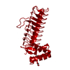
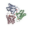
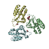
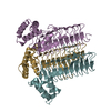
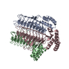
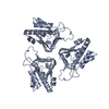
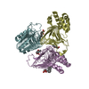
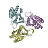
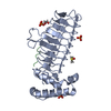
| Title | UDP N-ACETYLGLUCOSAMINE ACYLTRANSFERASE | ||||||
 Components Components | UDP N-ACETYLGLUCOSAMINE O-ACYLTRANSFERASE | ||||||
 Keywords Keywords | ACYLTRANSFERASE / TRANSFERASE / LIPID A BIOSYNTHESIS / LIPID SYNTHESIS | ||||||
| Function / homology |  Function and homology information Function and homology informationacyl-[acyl-carrier-protein]-UDP-N-acetylglucosamine O-acyltransferase / acyl-[acyl-carrier-protein]-UDP-N-acetylglucosamine O-acyltransferase activity / lipopolysaccharide biosynthetic process / lipid A biosynthetic process / membrane / identical protein binding / cytoplasm / cytosol Similarity search - Function | ||||||
| Biological species |  | ||||||
| Method |  X-RAY DIFFRACTION / Resolution: 2.6 Å X-RAY DIFFRACTION / Resolution: 2.6 Å | ||||||
 Authors Authors | Roderick, S.L. | ||||||
 Citation Citation | Journal: Science / Year: 1995 Title: A left-handed parallel beta helix in the structure of UDP-N-acetylglucosamine acyltransferase. Authors: Raetz, C.R. / Roderick, S.L. #1: Journal: Proteins / Year: 1995Title: Crystallization of Udp N-Acetylglucosamine O-Acyltransferase from Escherichia Coli Authors: Pfitzner, U. / Raetz, C.R.H. / Roderick, S.L. | ||||||
| History |
| ||||||
| Remark 650 | HELIX DETERMINATION METHOD: AUTHOR DETERMINED | ||||||
| Remark 700 | SHEET DETERMINATION METHOD: AUTHOR DETERMINED |
- Structure visualization
| Structure viewer | Molecule: MolmilJmol/JSmol |
|---|
- Downloads & links
-Download
| PDBx/mmCIF format | 1lxa.cif.gz | 59.1 KB | Display | PDBx/mmCIF format |
|---|---|---|---|---|
| PDB format | pdb1lxa.ent.gz | 44.4 KB | Display | PDB format |
| PDBx/mmJSON format | 1lxa.json.gz | Tree view | PDBx/mmJSON format | |
| Others |  Other downloads Other downloads |
-Validation report
| Arichive directory | https://data.pdbj.org/pub/pdb/validation_reports/lx/1lxaftp://data.pdbj.org/pub/pdb/validation_reports/lx/1lxa | HTTPS FTP |
|---|
-Related structure data
| Similar structure data |
|---|

-Links
 PDBj
PDBj


- Assembly
| Deposited unit | 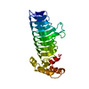
| ||||||||
|---|---|---|---|---|---|---|---|---|---|
| 1 | 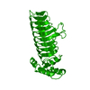
| ||||||||
| Unit cell |
|
-Components
| #1: Protein | Mass: 28117.018 Da / Num. of mol.: 1 / Mutation: S64Q, V65F, D125H Source method: isolated from a genetically manipulated source Source: (gene. exp.) References: UniProt: P0A722, acyl-[acyl-carrier-protein]-UDP-N-acetylglucosamine O-acyltransferase |
|---|---|
| Compound details | MET 1 THROUGH ILE 186 COMPRISES THE LEFT-HANDED BETA-HELICAL DOMAIN WITH TWO LOOP EXCURSIONS |
-Experimental details
-Experiment
| Experiment | Method: X-RAY DIFFRACTION / Number of used crystals: 1 |
|---|
- Sample preparation
| Crystal | Density Matthews: 2.87 Å3/Da / Density % sol: 57.21 % | ||||||||||||||||||
|---|---|---|---|---|---|---|---|---|---|---|---|---|---|---|---|---|---|---|---|
| Crystal | *PLUS Density % sol: 57 % | ||||||||||||||||||
| Crystal grow | *PLUS Method: vapor diffusion, hanging drop / PH range low: 6.8 / PH range high: 6.2 | ||||||||||||||||||
| Components of the solutions | *PLUS
|
-Data collection
| Diffraction source | Wavelength: 1.5418 Å |
|---|---|
| Detector | Type: SIEMENS / Detector: AREA DETECTOR / Date: 1995 |
| Radiation | Monochromatic (M) / Laue (L): M / Scattering type: x-ray |
| Radiation wavelength | Wavelength: 1.5418 Å / Relative weight: 1 |
| Reflection | Resolution: 2.6→99 Å / Num. obs: 8868 / % possible obs: 87 % / Observed criterion σ(I): 0 / Redundancy: 10 % / Rmerge(I) obs: 0.08 |
| Reflection | *PLUS Num. measured all: 79727 / Rmerge(I) obs: 0.08 |
- Processing
| Software |
| ||||||||||||||||||||
|---|---|---|---|---|---|---|---|---|---|---|---|---|---|---|---|---|---|---|---|---|---|
| Refinement | Resolution: 2.6→99 Å / Isotropic thermal model: KNOWLEDGE-BASED / σ(F): 0 / Stereochemistry target values: TNT SUPPLIED
| ||||||||||||||||||||
| Solvent computation | Solvent model: TWO PARAMETER RECIPROCAL SPACE / Bsol: 250 Å2 / ksol: 0.874 e/Å3 | ||||||||||||||||||||
| Refinement step | Cycle: LAST / Resolution: 2.6→99 Å
| ||||||||||||||||||||
| Refine LS restraints |
| ||||||||||||||||||||
| Software | *PLUS Name: X-PLOR/TNT / Classification: refinement | ||||||||||||||||||||
| Refinement | *PLUS Rfactor all: 0.184 | ||||||||||||||||||||
| Solvent computation | *PLUS | ||||||||||||||||||||
| Displacement parameters | *PLUS |
