 Movie
Movie Controller


+ Open data

- Basic information
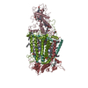
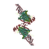
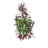
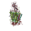
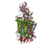
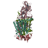
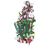
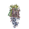
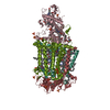
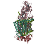
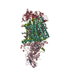
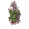
| Entry | Database: PDB / ID: 2jbl | ||||||||||||
|---|---|---|---|---|---|---|---|---|---|---|---|---|---|
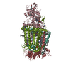
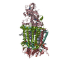
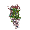
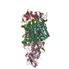
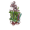
| Title | PHOTOSYNTHETIC REACTION CENTER FROM BLASTOCHLORIS VIRIDIS | ||||||||||||
 Components Components |
| ||||||||||||
 Keywords Keywords | ELECTRON TRANSPORT / CHROMOPHORE / FORMYLATION / CHLOROPHYLL / LIPOPROTEIN / STIGMATELLIN / METAL-BINDING / TRANSMEMBRANE / IRON / HEME / MEMBRANE / TRANSPORT / MAGNESIUM / PHOTOSYNTHESIS / REACTION CENTER / BACTERIOCHLOROPHYLL / PHOTOSYNTHETIC REACTION CENTER | ||||||||||||
| Function / homology |  Function and homology information Function and homology informationplasma membrane-derived chromatophore membrane / plasma membrane light-harvesting complex / bacteriochlorophyll binding / photosynthetic electron transport in photosystem II / photosynthesis, light reaction / photosynthesis / electron transfer activity / iron ion binding / heme binding / metal ion binding Similarity search - Function | ||||||||||||
| Biological species |  BLASTOCHLORIS VIRIDIS (bacteria) BLASTOCHLORIS VIRIDIS (bacteria) | ||||||||||||
| Method |  X-RAY DIFFRACTION / SYNCHROTRON / MOLECULAR REPLACEMENT / Resolution: 2.4 Å X-RAY DIFFRACTION / SYNCHROTRON / MOLECULAR REPLACEMENT / Resolution: 2.4 Å | ||||||||||||
 Authors Authors | Lancaster, C.R.D. | ||||||||||||
 Citation Citation | Journal: J.Mol.Biol. / Year: 2007 Title: A Comparison of Stigmatellin Conformations, Free and Bound to the Photosynthetic Reaction Center and the Cytochrome Bc(1) Complex. Authors: Lancaster, C.R.D. / Hunte, C. / Kelley, J. / Trumpower, B.L. / Ditchfield, R. #1: Journal: J.Biol.Chem. / Year: 2000Title: Structural Basis of the Drastically Increased Initial Electron Transfer Rate in the Reaction Center from a Rhodopseudomonas Viridis Mutant Described at 2.00-A Resolution Authors: Lancaster, C.R.D. / Bibikova, M. / Sabatino, P. / Oesterhelt, D. / Michel, H. #2: Journal: J.Mol.Biol. / Year: 1999Title: Refined Crystal Structures of Reaction Centres from Rhodopseudomonas Viridis in Complexes with the Herbicide Atrazine and Two Chiral Atrazine Derivatives Also Lead to a New Model of the Bound Carotenoid Authors: Lancaster, C.R. / Michel, H. #3: Journal: Biochim.Biophys.Acta / Year: 1998Title: Ubiquinone Reduction and Protonation in the Reaction Centre of Rhodopseudomonas Viridis: X-Ray Structures and Their Functional Implications Authors: Lancaster, C.R.D. #4: Journal: Structure / Year: 1997Title: The Coupling of Light-Induced Electron Transfer and Proton Uptake as Derived from Crystal Structures of Reaction Centres from Rhodopseudomonas Viridis Modified at the Binding Site of the Secondary Quinone, Qb Authors: Lancaster, C.R. / Michel, H. #5: Journal: J.Mol.Biol. / Year: 1995Title: Crystallographic Refinement at 2.3 A Resolution and Refined Model of the Photosynthetic Reaction Centre from Rhodopseudomonas Viridis Authors: Deisenhofer, J. / Epp, O. / Sinning, I. / Michel, H. #6: Journal: Science / Year: 1989 Title: The Photosynthetic Reaction Center from the Purple Bacterium Rhodopseudomonas Viridis Authors: Deisenhofer, J. / Michel, H. #7: Journal: Nature / Year: 1985 Title: Structure of the Protein Subunits in the Photosynthetic Reaction Centre of Rhodopseudomonas Viridis at 3 Angstroms Resolution Authors: Deisenhofer, J. / Epp, O. / Miki, K. / Huber, R. / Michel, H. #8: Journal: J.Mol.Biol. / Year: 1984 Title: X-Ray Structure Analysis of a Membrane Protein Complex. Electron Density Map at 3 A Resolution and a Model of the Chromophores of the Photosynthetic Reaction Center from Rhodopseudomonas Viridis Authors: Deisenhofer, J. / Epp, O. / Miki, K. / Huber, R. / Michel, H. #9: Journal: J.Mol.Biol. / Year: 1982 Title: Three-Dimensional Crystals of a Membrane Protein Complex. The Photosynthetic Reaction Centre from Rhodopseudomonas Viridis Authors: Michel, H. | ||||||||||||
| History |
| ||||||||||||
| Remark 700 | SHEET THE SHEET STRUCTURE OF THIS MOLECULE IS BIFURCATED. IN ORDER TO REPRESENT THIS FEATURE IN ... SHEET THE SHEET STRUCTURE OF THIS MOLECULE IS BIFURCATED. IN ORDER TO REPRESENT THIS FEATURE IN THE SHEET RECORDS BELOW, TWO SHEETS ARE DEFINED. |
- Structure visualization
| Structure viewer | Molecule: MolmilJmol/JSmol |
|---|
- Downloads & links
-Download
| PDBx/mmCIF format | 2jbl.cif.gz | 287.5 KB | Display | PDBx/mmCIF format |
|---|---|---|---|---|
| PDB format | pdb2jbl.ent.gz | 224.2 KB | Display | PDB format |
| PDBx/mmJSON format | 2jbl.json.gz | Tree view | PDBx/mmJSON format | |
| Others |  Other downloads Other downloads |
-Validation report
| Arichive directory | https://data.pdbj.org/pub/pdb/validation_reports/jb/2jblftp://data.pdbj.org/pub/pdb/validation_reports/jb/2jbl | HTTPS FTP |
|---|
-Related structure data
| Related structure data |  2ibzC  4prc S: Starting model for refinement C: citing same article ( |
|---|---|
| Similar structure data |


-Links
 PDBj
PDBj


- Assembly
| Deposited unit | 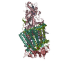
| ||||||||
|---|---|---|---|---|---|---|---|---|---|
| 1 |
| ||||||||
| Unit cell |
|
-Components
-Protein , 1 types, 1 molecules C
| #1: Protein | Mass: 39419.176 Da / Num. of mol.: 1 / Source method: isolated from a natural source / Details: GERMAN COLLECTION OF MICROORGANISMS (DSM 133) / Source: (natural) BLASTOCHLORIS VIRIDIS (bacteria) / References: UniProt: P07173 |
|---|
-REACTION CENTER PROTEIN ... , 3 types, 3 molecules HLM
| #2: Protein | Mass: 28557.453 Da / Num. of mol.: 1 / Source method: isolated from a natural source / Details: GERMAN COLLECTION OF MICROORGANISMS (DSM 133) / Source: (natural) BLASTOCHLORIS VIRIDIS (bacteria) / References: UniProt: P06008 |
|---|---|
| #3: Protein | Mass: 30469.104 Da / Num. of mol.: 1 / Source method: isolated from a natural source / Details: GERMAN COLLECTION OF MICROORGANISMS (DSM 133) / Source: (natural) BLASTOCHLORIS VIRIDIS (bacteria) / References: UniProt: P06009 |
| #4: Protein | Mass: 35932.188 Da / Num. of mol.: 1 / Source method: isolated from a natural source / Details: GERMAN COLLECTION OF MICROORGANISMS (DSM 133) / Source: (natural) BLASTOCHLORIS VIRIDIS (bacteria) / References: UniProt: P06010 |
-Non-polymers , 10 types, 366 molecules 


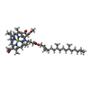
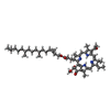
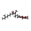

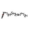
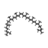

| #5: Chemical | ChemComp-HEC /  Mass: 618.503 Da / Num. of mol.: 4 / Source method: obtained synthetically / Formula: C34H34FeN4O4 Mass: 618.503 Da / Num. of mol.: 4 / Source method: obtained synthetically / Formula: C34H34FeN4O4#6: Chemical | ChemComp-LDA /  Mass: 229.402 Da / Num. of mol.: 6 / Source method: obtained synthetically / Formula: C14H31NO / Comment: LDAO, detergent*YM Mass: 229.402 Da / Num. of mol.: 6 / Source method: obtained synthetically / Formula: C14H31NO / Comment: LDAO, detergent*YM#7: Chemical | ChemComp-SO4 /  Mass: 96.063 Da / Num. of mol.: 4 / Source method: obtained synthetically / Formula: SO4 Mass: 96.063 Da / Num. of mol.: 4 / Source method: obtained synthetically / Formula: SO4#8: Chemical | ChemComp-BCB /  Mass: 909.488 Da / Num. of mol.: 4 / Source method: obtained synthetically / Formula: C55H72MgN4O6 Mass: 909.488 Da / Num. of mol.: 4 / Source method: obtained synthetically / Formula: C55H72MgN4O6#9: Chemical |  Mass: 887.199 Da / Num. of mol.: 2 / Source method: obtained synthetically / Formula: C55H74N4O6 Mass: 887.199 Da / Num. of mol.: 2 / Source method: obtained synthetically / Formula: C55H74N4O6#10: Chemical | ChemComp-SMA / |  Mass: 514.650 Da / Num. of mol.: 1 / Source method: obtained synthetically / Formula: C30H42O7 Mass: 514.650 Da / Num. of mol.: 1 / Source method: obtained synthetically / Formula: C30H42O7#11: Chemical | ChemComp-FE / |  Mass: 55.845 Da / Num. of mol.: 1 / Source method: obtained synthetically / Formula: Fe Mass: 55.845 Da / Num. of mol.: 1 / Source method: obtained synthetically / Formula: Fe#12: Chemical | ChemComp-MQ7 / |  Mass: 648.999 Da / Num. of mol.: 1 / Source method: obtained synthetically / Formula: C46H64O2 Mass: 648.999 Da / Num. of mol.: 1 / Source method: obtained synthetically / Formula: C46H64O2#13: Chemical | ChemComp-NS5 / |  Mass: 540.904 Da / Num. of mol.: 1 / Source method: obtained synthetically / Formula: C40H60 Mass: 540.904 Da / Num. of mol.: 1 / Source method: obtained synthetically / Formula: C40H60#14: Water | ChemComp-HOH / | Mass: 18.015 Da / Num. of mol.: 342 / Source method: isolated from a natural source / Formula: H2O |
|---|
-Details
| Has protein modification | Y |
|---|
-Experimental details
-Experiment
| Experiment | Method: X-RAY DIFFRACTION / Number of used crystals: 5 |
|---|
- Sample preparation
| Crystal | Density Matthews: 4.82 Å3/Da / Density % sol: 74.28 % |
|---|---|
| Crystal grow | pH: 6 / Details: pH 6.00 |
-Data collection
| Diffraction | Mean temperature: 263 K |
|---|---|
| Diffraction source | Source: SYNCHROTRON / Site: EMBL/DESY, HAMBURG  / Beamline: X11 / Wavelength: 0.92 / Beamline: X11 / Wavelength: 0.92 |
| Detector | Type: MARRESEARCH / Detector: IMAGE PLATE / Date: Nov 13, 1992 / Details: FOCUSING MIRROR |
| Radiation | Monochromator: BENT SINGLE-CRYSTAL GERMANIUM TRIANGULAR MONOCHROMATOR Protocol: SINGLE WAVELENGTH / Monochromatic (M) / Laue (L): M / Scattering type: x-ray |
| Radiation wavelength | Wavelength: 0.92 Å / Relative weight: 1 |
| Reflection | Resolution: 2.4→26.56 Å / Num. obs: 103499 / % possible obs: 92.3 % / Observed criterion σ(I): -3 / Redundancy: 1.9 % / Biso Wilson estimate: 28.1 Å2 / Rmerge(I) obs: 0.09 / Net I/σ(I): 6.9 |
| Reflection shell | Resolution: 2.4→2.46 Å / Redundancy: 1.4 % / Rmerge(I) obs: 0.31 / Mean I/σ(I) obs: 1.6 / % possible all: 86.2 |
- Processing
| Software |
| ||||||||||||||||||||||||||||||||||||||||||||||||||||||||||||
|---|---|---|---|---|---|---|---|---|---|---|---|---|---|---|---|---|---|---|---|---|---|---|---|---|---|---|---|---|---|---|---|---|---|---|---|---|---|---|---|---|---|---|---|---|---|---|---|---|---|---|---|---|---|---|---|---|---|---|---|---|---|
| Refinement | Method to determine structure: MOLECULAR REPLACEMENT Starting model: PDB ENTRY 4PRC 4prc Resolution: 2.4→26.56 Å / Rfactor Rfree error: 0.005 / Data cutoff high absF: 3307055.41 / Isotropic thermal model: RESTRAINED / Cross valid method: THROUGHOUT / σ(F): 0 / Stereochemistry target values: MLF
| ||||||||||||||||||||||||||||||||||||||||||||||||||||||||||||
| Solvent computation | Solvent model: FLAT MODEL / Bsol: 56.1587 Å2 / ksol: 0.285982 e/Å3 | ||||||||||||||||||||||||||||||||||||||||||||||||||||||||||||
| Displacement parameters | Biso mean: 37.7 Å2
| ||||||||||||||||||||||||||||||||||||||||||||||||||||||||||||
| Refine analyze |
| ||||||||||||||||||||||||||||||||||||||||||||||||||||||||||||
| Refinement step | Cycle: LAST / Resolution: 2.4→26.56 Å
| ||||||||||||||||||||||||||||||||||||||||||||||||||||||||||||
| Refine LS restraints |
| ||||||||||||||||||||||||||||||||||||||||||||||||||||||||||||
| LS refinement shell | Resolution: 2.4→2.55 Å / Rfactor Rfree error: 0.02 / Total num. of bins used: 6
| ||||||||||||||||||||||||||||||||||||||||||||||||||||||||||||
| Xplor file |
|
