 Movie
Movie Controller

[English] 日本語
 Yorodumi
- PDB-1prc: CRYSTALLOGRAPHIC REFINEMENT AT 2.3 ANGSTROMS RESOLUTION AND REFIN... -
+ Open data

- Basic information
| Entry | Database: PDB / ID: 1prc | |||||||||
|---|---|---|---|---|---|---|---|---|---|---|
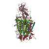
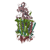
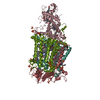
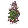
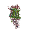
| Title | CRYSTALLOGRAPHIC REFINEMENT AT 2.3 ANGSTROMS RESOLUTION AND REFINED MODEL OF THE PHOTOSYNTHETIC REACTION CENTER FROM RHODOPSEUDOMONAS VIRIDIS | |||||||||
 Components Components | (PHOTOSYNTHETIC REACTION ...) x 4 | |||||||||
 Keywords Keywords | PHOTOSYNTHETIC REACTION CENTER | |||||||||
| Function / homology |  Function and homology information Function and homology informationplasma membrane-derived chromatophore membrane / plasma membrane light-harvesting complex / bacteriochlorophyll binding / photosynthetic electron transport in photosystem II / photosynthesis, light reaction / photosynthesis / electron transfer activity / iron ion binding / heme binding / metal ion binding Similarity search - Function | |||||||||
| Biological species |  Blastochloris viridis (bacteria) Blastochloris viridis (bacteria) | |||||||||
| Method |  X-RAY DIFFRACTION / Resolution: 2.3 Å X-RAY DIFFRACTION / Resolution: 2.3 Å | |||||||||
 Authors Authors | Deisenhofer, J. / Epp, O. / Miki, K. / Huber, R. / Michel, H. | |||||||||
 Citation Citation | Journal: J.Mol.Biol. / Year: 1995 Title: Crystallographic refinement at 2.3 A resolution and refined model of the photosynthetic reaction centre from Rhodopseudomonas viridis. Authors: Deisenhofer, J. / Epp, O. / Sinning, I. / Michel, H. #1: Journal: Science / Year: 1989Title: The Photosynthetic Reaction Center from the Purple Bacterium Rhodopseudomonas Viridis Authors: Deisenhofer, J. / Michel, H. #2: Journal: Nature / Year: 1985Title: Structure of the Protein Subunits in the Photosynthetic Reaction Centre of Rhodopseudomonas Viridis at 3 Angstroms Resolution Authors: Deisenhofer, J. / Epp, O. / Miki, K. / Huber, R. / Michel, H. #3: Journal: J.Mol.Biol. / Year: 1984Title: X-Ray Structure Analysis of a Membrane Protein Complex. Electron Density Map at 3 Angstroms Resolution and a Model of the Chromophores of the Photosynthetic Reaction Center from Rhodopseudomonas Viridis Authors: Deisenhofer, J. / Epp, O. / Miki, K. / Huber, R. / Michel, H. #4: Journal: J.Mol.Biol. / Year: 1982Title: Three-Dimensional Crystals of a Membrane Protein Complex. The Photosynthetic Reaction Centre from Rhodopseudomonas Viridis Authors: Michel, H. | |||||||||
| History |
|
- Structure visualization
| Structure viewer | Molecule: MolmilJmol/JSmol |
|---|
- Downloads & links
-Download
| PDBx/mmCIF format | 1prc.cif.gz | 280.5 KB | Display | PDBx/mmCIF format |
|---|---|---|---|---|
| PDB format | pdb1prc.ent.gz | 218.9 KB | Display | PDB format |
| PDBx/mmJSON format | 1prc.json.gz | Tree view | PDBx/mmJSON format | |
| Others |  Other downloads Other downloads |
-Validation report
| Arichive directory | https://data.pdbj.org/pub/pdb/validation_reports/pr/1prcftp://data.pdbj.org/pub/pdb/validation_reports/pr/1prc | HTTPS FTP |
|---|
-Related structure data
| Similar structure data |
|---|

-Links
 PDBj
PDBj


- Assembly
| Deposited unit | 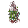
| ||||||||
|---|---|---|---|---|---|---|---|---|---|
| 1 |
| ||||||||
| 2 | 
| ||||||||
| Unit cell |
| ||||||||
| Atom site foot note | 1: RESIDUES PRO C 6, PRO C 153, PRO C 330, PRO M 48, PRO H 42, AND PRO H 79 ARE CIS PROLINES. 2: SEE REMARK 4. | ||||||||
| Components on special symmetry positions |
|
-Components
-PHOTOSYNTHETIC REACTION ... , 4 types, 4 molecules CLMH
| #1: Protein | Mass: 37450.801 Da / Num. of mol.: 1 Source method: isolated from a genetically manipulated source Source: (gene. exp.) Blastochloris viridis (bacteria) / References: UniProt: P07173 |
|---|---|
| #2: Protein | Mass: 30469.104 Da / Num. of mol.: 1 Source method: isolated from a genetically manipulated source Source: (gene. exp.) Blastochloris viridis (bacteria) / References: UniProt: P06009 |
| #3: Protein | Mass: 35932.188 Da / Num. of mol.: 1 Source method: isolated from a genetically manipulated source Source: (gene. exp.) Blastochloris viridis (bacteria) / References: UniProt: P06010 |
| #4: Protein | Mass: 28557.453 Da / Num. of mol.: 1 Source method: isolated from a genetically manipulated source Source: (gene. exp.) Blastochloris viridis (bacteria) / References: UniProt: P06008 |
-Non-polymers , 10 types, 224 molecules 


| #5: Chemical | ChemComp-HEC /  Mass: 618.503 Da / Num. of mol.: 4 / Source method: obtained synthetically / Formula: C34H34FeN4O4 Mass: 618.503 Da / Num. of mol.: 4 / Source method: obtained synthetically / Formula: C34H34FeN4O4#6: Chemical | ChemComp-BCB /  Mass: 909.488 Da / Num. of mol.: 4 / Source method: obtained synthetically / Formula: C55H72MgN4O6 Mass: 909.488 Da / Num. of mol.: 4 / Source method: obtained synthetically / Formula: C55H72MgN4O6#7: Chemical |  Mass: 887.199 Da / Num. of mol.: 2 / Source method: obtained synthetically / Formula: C55H74N4O6 Mass: 887.199 Da / Num. of mol.: 2 / Source method: obtained synthetically / Formula: C55H74N4O6#8: Chemical | ChemComp-UQ1 / |  Mass: 250.290 Da / Num. of mol.: 1 / Source method: obtained synthetically / Formula: C14H18O4 Mass: 250.290 Da / Num. of mol.: 1 / Source method: obtained synthetically / Formula: C14H18O4#9: Chemical | ChemComp-FE / |  Mass: 55.845 Da / Num. of mol.: 1 / Source method: obtained synthetically / Formula: Fe Mass: 55.845 Da / Num. of mol.: 1 / Source method: obtained synthetically / Formula: Fe#10: Chemical | ChemComp-SO4 /  Mass: 96.063 Da / Num. of mol.: 7 / Source method: obtained synthetically / Formula: SO4 Mass: 96.063 Da / Num. of mol.: 7 / Source method: obtained synthetically / Formula: SO4#11: Chemical | ChemComp-MQ7 / |  Mass: 648.999 Da / Num. of mol.: 1 / Source method: obtained synthetically / Formula: C46H64O2 Mass: 648.999 Da / Num. of mol.: 1 / Source method: obtained synthetically / Formula: C46H64O2#12: Chemical | ChemComp-NS1 / |  Mass: 540.904 Da / Num. of mol.: 1 / Source method: obtained synthetically / Formula: C40H60 Mass: 540.904 Da / Num. of mol.: 1 / Source method: obtained synthetically / Formula: C40H60#13: Chemical |  Mass: 229.402 Da / Num. of mol.: 2 / Source method: obtained synthetically / Formula: C14H31NO / Comment: LDAO, detergent*YM Mass: 229.402 Da / Num. of mol.: 2 / Source method: obtained synthetically / Formula: C14H31NO / Comment: LDAO, detergent*YM#14: Water | ChemComp-HOH / | Mass: 18.015 Da / Num. of mol.: 201 / Source method: isolated from a natural source / Formula: H2O |
|---|
-Details
| Has protein modification | Y |
|---|
-Experimental details
-Experiment
| Experiment | Method: X-RAY DIFFRACTION |
|---|
- Sample preparation
| Crystal | Density Matthews: 5.36 Å3/Da / Density % sol: 77.04 % | |||||||||||||||
|---|---|---|---|---|---|---|---|---|---|---|---|---|---|---|---|---|
| Crystal grow | *PLUS pH: 6 / Method: vapor diffusion / Details: Michel, H., (1982) J.Mol.Biol., 158, 567. | |||||||||||||||
| Components of the solutions | *PLUS
|
-Data collection
| Radiation | Scattering type: x-ray |
|---|---|
| Radiation wavelength | Relative weight: 1 |
| Reflection | *PLUS Highest resolution: 2.3 Å / Num. obs: 95762 / Observed criterion σ(I): 2 / Rmerge(I) obs: 0.091 |
| Reflection shell | *PLUS Highest resolution: 2.3 Å / Lowest resolution: 2.4 Å / % possible obs: 49.2 % |
- Processing
| Software | Name: TNT / Classification: refinement | ||||||||||||
|---|---|---|---|---|---|---|---|---|---|---|---|---|---|
| Refinement | Resolution: 2.3→19.99 Å /
| ||||||||||||
| Refine analyze | Luzzati coordinate error obs: 0.26 Å | ||||||||||||
| Refinement step | Cycle: LAST / Resolution: 2.3→19.99 Å
| ||||||||||||
| Refinement | *PLUS Rfactor obs: 0.193 | ||||||||||||
| Solvent computation | *PLUS | ||||||||||||
| Displacement parameters | *PLUS Biso mean: 22.5 Å2 |
