 Movie
Movie Controller

[English] 日本語
 Yorodumi
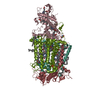
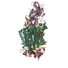
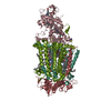
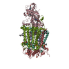
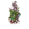
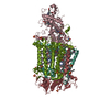
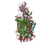
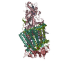
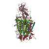
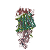
- PDB-3t6e: Crystal Structure of the Reaction Centre from Blastochloris virid... -
+ Open data

- Basic information
| Entry | Database: PDB / ID: 3t6e | ||||||
|---|---|---|---|---|---|---|---|
| Title | Crystal Structure of the Reaction Centre from Blastochloris viridis strain DSM 133 (ATCC 19567) substrain-94 | ||||||
 Components Components |
| ||||||
 Keywords Keywords | ELECTRON TRANSPORT / Pigment-protein complex / Electron transfer / Photosynthesis / Photosynthetic reaction center / Photosynthetic membranes / Evolution / Genetic drift | ||||||
| Function / homology |  Function and homology information Function and homology informationplasma membrane-derived chromatophore membrane / plasma membrane light-harvesting complex / bacteriochlorophyll binding / photosynthetic electron transport in photosystem II / photosynthesis, light reaction / photosynthesis / electron transfer activity / iron ion binding / heme binding / metal ion binding Similarity search - Function | ||||||
| Biological species |  Blastochloris viridis (bacteria) Blastochloris viridis (bacteria) | ||||||
| Method |  X-RAY DIFFRACTION / SYNCHROTRON / MOLECULAR REPLACEMENT / Resolution: 1.92 Å X-RAY DIFFRACTION / SYNCHROTRON / MOLECULAR REPLACEMENT / Resolution: 1.92 Å | ||||||
 Authors Authors | Roszak, A.W. / Gardiner, A.T. / Isaacs, N.W. / Cogdell, R.J. | ||||||
 Citation Citation | Journal: Biochem.J. / Year: 2012 Title: New insights into the structure of the reaction centre from Blastochloris viridis: evolution in the laboratory. Authors: Roszak, A.W. / Moulisova, V. / Reksodipuro, A.D. / Gardiner, A.T. / Fujii, R. / Hashimoto, H. / Isaacs, N.W. / Cogdell, R.J. | ||||||
| History |
|
- Structure visualization
| Structure viewer | Molecule: MolmilJmol/JSmol |
|---|
- Downloads & links
-Download
| PDBx/mmCIF format | 3t6e.cif.gz | 623 KB | Display | PDBx/mmCIF format |
|---|---|---|---|---|
| PDB format | pdb3t6e.ent.gz | 507 KB | Display | PDB format |
| PDBx/mmJSON format | 3t6e.json.gz | Tree view | PDBx/mmJSON format | |
| Others |  Other downloads Other downloads |
-Validation report
| Arichive directory | https://data.pdbj.org/pub/pdb/validation_reports/t6/3t6eftp://data.pdbj.org/pub/pdb/validation_reports/t6/3t6e | HTTPS FTP |
|---|
-Related structure data
| Related structure data |  3t6dSC S: Starting model for refinement C: citing same article ( |
|---|---|
| Similar structure data |


-Links
 PDBj
PDBj


- Assembly
| Deposited unit | 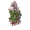
| ||||||||
|---|---|---|---|---|---|---|---|---|---|
| 1 |
| ||||||||
| Unit cell |
| ||||||||
| Components on special symmetry positions |
|
-Components
-Protein , 1 types, 1 molecules C
| #1: Protein | Mass: 39419.176 Da / Num. of mol.: 1 / Source method: isolated from a natural source Details: substrain-94 is identical at the amino acid level with the original strain DSM 133 Source: (natural) Blastochloris viridis (bacteria) / References: UniProt: P07173 |
|---|
-Reaction center protein ... , 3 types, 3 molecules HLM
| #2: Protein | Mass: 28557.453 Da / Num. of mol.: 1 / Source method: isolated from a natural source Details: substrain-94 is identical at the amino acid level with the original strain DSM 133 Source: (natural) Blastochloris viridis (bacteria) / References: UniProt: P06008 |
|---|---|
| #3: Protein | Mass: 30469.104 Da / Num. of mol.: 1 / Source method: isolated from a natural source Details: substrain-94 is identical at the amino acid level with the original strain DSM 133 Source: (natural) Blastochloris viridis (bacteria) / References: UniProt: P06009 |
| #4: Protein | Mass: 35948.188 Da / Num. of mol.: 1 / Source method: isolated from a natural source Details: substrain-94 is identical at the amino acid level with the original strain DSM 133 Source: (natural) Blastochloris viridis (bacteria) / References: UniProt: P06010 |
-Non-polymers , 13 types, 1084 molecules 

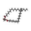

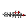

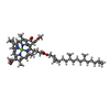
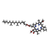
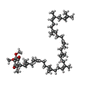


| #5: Chemical | ChemComp-HEC /  Mass: 618.503 Da / Num. of mol.: 4 / Source method: obtained synthetically / Formula: C34H34FeN4O4 Mass: 618.503 Da / Num. of mol.: 4 / Source method: obtained synthetically / Formula: C34H34FeN4O4#6: Chemical | ChemComp-LDA /  Mass: 229.402 Da / Num. of mol.: 16 / Source method: obtained synthetically / Formula: C14H31NO / Comment: LDAO, detergent*YM Mass: 229.402 Da / Num. of mol.: 16 / Source method: obtained synthetically / Formula: C14H31NO / Comment: LDAO, detergent*YM#7: Chemical | ChemComp-DGA /  Mass: 625.018 Da / Num. of mol.: 4 / Source method: obtained synthetically / Formula: C39H76O5 Mass: 625.018 Da / Num. of mol.: 4 / Source method: obtained synthetically / Formula: C39H76O5#8: Chemical | ChemComp-SO4 /  Mass: 96.063 Da / Num. of mol.: 26 / Source method: obtained synthetically / Formula: SO4 Mass: 96.063 Da / Num. of mol.: 26 / Source method: obtained synthetically / Formula: SO4#9: Chemical | ChemComp-HTO /  Mass: 148.200 Da / Num. of mol.: 8 / Source method: obtained synthetically / Formula: C7H16O3 Mass: 148.200 Da / Num. of mol.: 8 / Source method: obtained synthetically / Formula: C7H16O3#10: Chemical | ChemComp-GOL /  Mass: 92.094 Da / Num. of mol.: 41 / Source method: obtained synthetically / Formula: C3H8O3 Mass: 92.094 Da / Num. of mol.: 41 / Source method: obtained synthetically / Formula: C3H8O3#11: Chemical | ChemComp-BCB /  Mass: 909.488 Da / Num. of mol.: 4 / Source method: obtained synthetically / Formula: C55H72MgN4O6 Mass: 909.488 Da / Num. of mol.: 4 / Source method: obtained synthetically / Formula: C55H72MgN4O6#12: Chemical |  Mass: 887.199 Da / Num. of mol.: 2 / Source method: obtained synthetically / Formula: C55H74N4O6 Mass: 887.199 Da / Num. of mol.: 2 / Source method: obtained synthetically / Formula: C55H74N4O6#13: Chemical |  Mass: 795.226 Da / Num. of mol.: 2 / Source method: obtained synthetically / Formula: C54H82O4 Mass: 795.226 Da / Num. of mol.: 2 / Source method: obtained synthetically / Formula: C54H82O4#14: Chemical | ChemComp-MQ9 / |  Mass: 785.233 Da / Num. of mol.: 1 / Source method: obtained synthetically / Formula: C56H80O2 Mass: 785.233 Da / Num. of mol.: 1 / Source method: obtained synthetically / Formula: C56H80O2#15: Chemical | ChemComp-FE2 / |  Mass: 55.845 Da / Num. of mol.: 1 / Source method: obtained synthetically / Formula: Fe Mass: 55.845 Da / Num. of mol.: 1 / Source method: obtained synthetically / Formula: Fe#16: Chemical | ChemComp-NS5 / |  Mass: 540.904 Da / Num. of mol.: 1 / Source method: obtained synthetically / Formula: C40H60 Mass: 540.904 Da / Num. of mol.: 1 / Source method: obtained synthetically / Formula: C40H60#17: Water | ChemComp-HOH / | Mass: 18.015 Da / Num. of mol.: 974 / Source method: isolated from a natural source / Formula: H2O |
|---|
-Details
| Has protein modification | Y |
|---|
-Experimental details
-Experiment
| Experiment | Method: X-RAY DIFFRACTION / Number of used crystals: 1 |
|---|
- Sample preparation
| Crystal grow | Temperature: 292 K / Method: vapor diffusion, sitting drop / pH: 6 Details: Protein in 20 mM sodium phosphate buffer and 0.1% lauryldimethylamine N,N-oxide (LDAO) detergent, precipitant 1.5 M ammonium sulfate, amphiphile 3% heptanetriol, reservoir solution 2.2-2.4 M ...Details: Protein in 20 mM sodium phosphate buffer and 0.1% lauryldimethylamine N,N-oxide (LDAO) detergent, precipitant 1.5 M ammonium sulfate, amphiphile 3% heptanetriol, reservoir solution 2.2-2.4 M ammonium sulphate, pH 6.0, VAPOR DIFFUSION, SITTING DROP, temperature 292K |
|---|
-Data collection
| Diffraction | Mean temperature: 100 K |
|---|---|
| Diffraction source | Source: SYNCHROTRON / Site: Diamond  / Beamline: I02 / Wavelength: 0.9194 Å / Beamline: I02 / Wavelength: 0.9194 Å |
| Detector | Type: ADSC QUANTUM 315r / Detector: CCD / Date: May 8, 2010 / Details: Mirrors |
| Radiation | Monochromator: Si(111) crystal / Protocol: SINGLE WAVELENGTH / Monochromatic (M) / Laue (L): M / Scattering type: x-ray |
| Radiation wavelength | Wavelength: 0.9194 Å / Relative weight: 1 |
| Reflection | Resolution: 1.92→43.46 Å / Num. all: 212134 / Num. obs: 212134 / % possible obs: 99.5 % / Observed criterion σ(F): 0 / Observed criterion σ(I): 0 / Redundancy: 19.35 % / Biso Wilson estimate: 36 Å2 / Rmerge(I) obs: 0.115 / Rsym value: 0.119 / Net I/σ(I): 9.4 |
| Reflection shell | Resolution: 1.92→1.99 Å / Redundancy: 18.87 % / Rmerge(I) obs: 0.823 / Mean I/σ(I) obs: 1.6 / Num. unique all: 20895 / Rsym value: 0.843 / % possible all: 99.1 |
- Processing
| Software |
| ||||||||||||||||||||||||||||||||||||||||||||||||||||||||||||||||||||||
|---|---|---|---|---|---|---|---|---|---|---|---|---|---|---|---|---|---|---|---|---|---|---|---|---|---|---|---|---|---|---|---|---|---|---|---|---|---|---|---|---|---|---|---|---|---|---|---|---|---|---|---|---|---|---|---|---|---|---|---|---|---|---|---|---|---|---|---|---|---|---|---|
| Refinement | Method to determine structure: MOLECULAR REPLACEMENT Starting model: PDB entry 3T6D Resolution: 1.92→43.46 Å / Cor.coef. Fo:Fc: 0.972 / Cor.coef. Fo:Fc free: 0.965 / SU B: 4.678 / SU ML: 0.059 / Isotropic thermal model: Isotropic / Cross valid method: THROUGHOUT / σ(F): 0 / ESU R Free: 0.085 / Stereochemistry target values: MAXIMUM LIKELIHOOD / Details: HYDROGENS HAVE BEEN ADDED IN RIDING POSITIONS
| ||||||||||||||||||||||||||||||||||||||||||||||||||||||||||||||||||||||
| Solvent computation | Ion probe radii: 0.8 Å / Shrinkage radii: 0.8 Å / VDW probe radii: 1.2 Å / Solvent model: BABINET MODEL WITH MASK | ||||||||||||||||||||||||||||||||||||||||||||||||||||||||||||||||||||||
| Displacement parameters | Biso mean: 46.86 Å2
| ||||||||||||||||||||||||||||||||||||||||||||||||||||||||||||||||||||||
| Refinement step | Cycle: LAST / Resolution: 1.92→43.46 Å
| ||||||||||||||||||||||||||||||||||||||||||||||||||||||||||||||||||||||
| Refine LS restraints |
| ||||||||||||||||||||||||||||||||||||||||||||||||||||||||||||||||||||||
| LS refinement shell | Resolution: 1.92→1.97 Å / Total num. of bins used: 20
| ||||||||||||||||||||||||||||||||||||||||||||||||||||||||||||||||||||||
| Refinement TLS params. | Method: refined / Origin x: -0.595 Å / Origin y: -49.212 Å / Origin z: 10.558 Å
| ||||||||||||||||||||||||||||||||||||||||||||||||||||||||||||||||||||||
| Refinement TLS group |
|
