[English] 日本語
Yorodumi
- PDB-2iyl: Structure of an FtsY:GDP complex -

+
Open data


ID or keywords:

Loading...

-
Basic information

Entry
Database: PDB / ID: 2iyl
TitleStructure of an FtsY:GDP complex
ComponentsCELL DIVISION PROTEIN FTSY
KeywordsCELL CYCLE / INNER MEMBRANE / RIBONUCLEOPROTEIN / NUCLEOTIDE-BINDING / SIGNAL RECOGNITION PARTICLE / SRP / GDP / FFH / FTSY / GTPASE / RNA-BINDING / GTP-BINDING / CELL DIVISION / MEMBRANE
Function / homology
Function and homology information


signal-recognition-particle GTPase / SRP-dependent cotranslational protein targeting to membrane / GTPase activity / GTP binding / ATP hydrolysis activity / plasma membrane / cytoplasm
Similarity search - Function
SRP54, nucleotide-binding domain / Signal-recognition particle receptor FtsY / SRP/SRP receptor, N-terminal / SRP54-type proteins GTP-binding domain signature. / Signal recognition particle SRP54, helical bundle / Signal recognition particle SRP54, N-terminal domain superfamily / SRP54-type protein, helical bundle domain / SRP54-type protein, helical bundle domain / Signal recognition particle, SRP54 subunit, GTPase domain / SRP54-type protein, GTPase domain ...SRP54, nucleotide-binding domain / Signal-recognition particle receptor FtsY / SRP/SRP receptor, N-terminal / SRP54-type proteins GTP-binding domain signature. / Signal recognition particle SRP54, helical bundle / Signal recognition particle SRP54, N-terminal domain superfamily / SRP54-type protein, helical bundle domain / SRP54-type protein, helical bundle domain / Signal recognition particle, SRP54 subunit, GTPase domain / SRP54-type protein, GTPase domain / SRP54-type protein, GTPase domain / Four Helix Bundle (Hemerythrin (Met), subunit A) / P-loop containing nucleotide triphosphate hydrolases / ATPases associated with a variety of cellular activities / AAA+ ATPase domain / Up-down Bundle / P-loop containing nucleoside triphosphate hydrolase / Rossmann fold / 3-Layer(aba) Sandwich / Mainly Alpha / Alpha Beta
Similarity search - Domain/homology
GUANOSINE-5'-DIPHOSPHATE / Signal recognition particle receptor FtsY
Similarity search - Component
Biological speciesTHERMUS AQUATICUS (bacteria)
MethodX-RAY DIFFRACTION / SYNCHROTRON / MOLECULAR REPLACEMENT / Resolution: 2.1 Å
AuthorsFocia, P.J. / Freymann, D.M.
CitationJournal: Proteins / Year: 2007
Title: X-ray structure of the T. aquaticus FtsY:GDP complex suggests functional roles for the C-terminal helix of the SRP GTPases.
Authors: Gawronski-Salerno, J. / Coon 5th., J.S. / Focia, P.J. / Freymann, D.M.
History
DepositionJul 18, 2006Deposition site: PDBE / Processing site: PDBE
Revision 1.0Jan 2, 2007Provider: repository / Type: Initial release
Revision 1.1May 8, 2011Group: Version format compliance
Revision 1.2Jul 13, 2011Group: Version format compliance
Revision 2.0Dec 19, 2018Group: Atomic model / Data collection ...Atomic model / Data collection / Database references / Derived calculations / Source and taxonomy
Category: atom_site / citation ...atom_site / citation / citation_author / entity_src_gen / struct_conn
Item: _atom_site.label_alt_id / _citation.journal_id_ISSN ..._atom_site.label_alt_id / _citation.journal_id_ISSN / _citation.page_last / _citation.pdbx_database_id_DOI / _citation.title / _citation_author.name / _entity_src_gen.pdbx_host_org_ncbi_taxonomy_id / _entity_src_gen.pdbx_host_org_scientific_name / _entity_src_gen.pdbx_host_org_strain / _entity_src_gen.pdbx_host_org_variant
Revision 2.1Dec 13, 2023Group: Data collection / Database references ...Data collection / Database references / Other / Refinement description
Category: chem_comp_atom / chem_comp_bond ...chem_comp_atom / chem_comp_bond / database_2 / pdbx_database_status / pdbx_initial_refinement_model
Item: _database_2.pdbx_DOI / _database_2.pdbx_database_accession / _pdbx_database_status.status_code_sf

-
Structure visualization

Structure viewerMolecule:
MolmilJmol/JSmol

Downloads & links

-
Assembly

Deposited unit
D: CELL DIVISION PROTEIN FTSY
hetero molecules


Theoretical massNumber of molelcules
Total (without water)31,4004
Polymers30,7651
Non-polymers6353
Water2,594144
1


  • Idetical with deposited unit
  • defined by author&software
TypeNameSymmetry operationNumber
identity operation1_555x,y,z1
MethodPQS
Unit cell
Length a, b, c (Å)105.196, 105.196, 54.500
Angle α, β, γ (deg.)90.00, 90.00, 90.00
Int Tables number79
Space group name H-MI4

-
Components

#1: Protein CELL DIVISION PROTEIN FTSY / / FTSY


Mass: 30764.586 Da / Num. of mol.: 1 / Fragment: NGD20, RESIDUES 20-303
Source method: isolated from a genetically manipulated source
Source: (gene. exp.) THERMUS AQUATICUS (bacteria) / Plasmid: PJGS3 / Production host: ESCHERICHIA COLI BL21(DE3) (bacteria) / Variant (production host): ROSETTA-2 pLysS / References: UniProt: P83749
#2: Chemical ChemComp-GDP / GUANOSINE-5'-DIPHOSPHATE / Guanosine diphosphate


Type: RNA linking / Mass: 443.201 Da / Num. of mol.: 1 / Source method: obtained synthetically / Formula: C10H15N5O11P2 / Comment: GDP, energy-carrying molecule*YM
#3: Chemical ChemComp-SO4 / SULFATE ION / Sulfate


Mass: 96.063 Da / Num. of mol.: 2 / Source method: obtained synthetically / Formula: SO4
#4: Water ChemComp-HOH / water / Water


Mass: 18.015 Da / Num. of mol.: 144 / Source method: isolated from a natural source / Formula: H2O
Sequence detailsRESIDUES 2-20 DELETED IN THE FTSY NG DOMAIN EXPRESSION CONSTRUCT FTSY NGD20. MET 1 NOT PRESENT.

-
Experimental details

-
Experiment

ExperimentMethod: X-RAY DIFFRACTION / Number of used crystals: 1

-
Sample preparation

Crystal
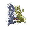
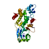
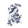
Density Matthews: 2.39 Å3/Da / Density % sol: 48.06 %
Description: CHAIN D OF ENTRY 1OKK, THE FTSY NG DOMAIN IN ITS HETERODIMERIC COMPLEX WITH FFH NG, WAS USED FOR MOLECULAR REPLACEMENT

-
Data collection

DiffractionMean temperature: 100 K
Diffraction sourceSource: SYNCHROTRON / Site: APS / Beamline: 14-BM-C / Wavelength: 0.9
DetectorType: ADSC CCD / Detector: CCD / Date: Dec 17, 2004
RadiationProtocol: SINGLE WAVELENGTH / Monochromatic (M) / Laue (L): M / Scattering type: x-ray
Radiation wavelengthWavelength: 0.9 Å / Relative weight: 1
ReflectionResolution: 2.06→48.39 Å / Num. obs: 136283 / % possible obs: 99.4 % / Observed criterion σ(I): 0 / Redundancy: 7.4 % / Rmerge(I) obs: 0.08 / Net I/σ(I): 20.1
Reflection shellResolution: 2.06→2.17 Å / Redundancy: 7.7 % / Rmerge(I) obs: 0.44 / Mean I/σ(I) obs: 3.9 / % possible all: 100

-
Processing

Software
NameVersionClassification
REFMAC5.2.0005refinement
MOSFLMdata reduction
SCALAdata scaling
PHASERphasing
RefinementMethod to determine structure: MOLECULAR REPLACEMENT
Starting model: PDB ENTRY 1OKK
Resolution: 2.1→11.87 Å / Cor.coef. Fo:Fc: 0.945 / Cor.coef. Fo:Fc free: 0.878 / SU B: 5.455 / SU ML: 0.15 / Cross valid method: THROUGHOUT / ESU R: 0.244 / ESU R Free: 0.231 / Stereochemistry target values: MAXIMUM LIKELIHOOD
Details: HYDROGENS HAVE BEEN ADDED IN THE RIDING POSITIONS. DISORDERED REGIONS WERE OMITTED FROM THE MODEL. THESE INCLUDE RESIDUES 80-82, 95-96, 141-146, AND 235- 236. THREE GLUTAMATE SIDECHAINS, ...Details: HYDROGENS HAVE BEEN ADDED IN THE RIDING POSITIONS. DISORDERED REGIONS WERE OMITTED FROM THE MODEL. THESE INCLUDE RESIDUES 80-82, 95-96, 141-146, AND 235- 236. THREE GLUTAMATE SIDECHAINS, D70, D151, AND D295, EXHIBIT EVIDENCE OF RADIATION DAMAGE AND THEIR CARBOXYLATE GROUP CD- OE1-OE2 ATOMS ARE MODELED AT 0.5 OCCUPANCY.
RfactorNum. reflection% reflectionSelection details
Rfree0.291 895 5.2 %RANDOM
Rwork0.207 ---
obs0.211 16465 99.5 %-
Solvent computationIon probe radii: 0.8 Å / Shrinkage radii: 0.8 Å / VDW probe radii: 1.2 Å / Solvent model: BABINET MODEL WITH MASK
Displacement parametersBiso mean: 44.96 Å2
Baniso -1Baniso -2Baniso -3
1-0 Å20 Å20 Å2
2--0 Å20 Å2
3----0.01 Å2
Refinement stepCycle: LAST / Resolution: 2.1→11.87 Å
ProteinNucleic acidLigandSolventTotal
Num. atoms2063 0 38 144 2245
Refine LS restraints
Refine-IDTypeDev idealDev ideal targetNumber
X-RAY DIFFRACTIONr_bond_refined_d0.0140.0222143
X-RAY DIFFRACTIONr_bond_other_d
X-RAY DIFFRACTIONr_angle_refined_deg1.5212.0192901
X-RAY DIFFRACTIONr_angle_other_deg
X-RAY DIFFRACTIONr_dihedral_angle_1_deg5.5775268
X-RAY DIFFRACTIONr_dihedral_angle_2_deg35.17825.24482
X-RAY DIFFRACTIONr_dihedral_angle_3_deg17.82215388
X-RAY DIFFRACTIONr_dihedral_angle_4_deg11.1551511
X-RAY DIFFRACTIONr_chiral_restr0.10.2332
X-RAY DIFFRACTIONr_gen_planes_refined0.0050.021540
X-RAY DIFFRACTIONr_gen_planes_other
X-RAY DIFFRACTIONr_nbd_refined0.2170.21035
X-RAY DIFFRACTIONr_nbd_other
X-RAY DIFFRACTIONr_nbtor_refined0.3060.21445
X-RAY DIFFRACTIONr_nbtor_other
X-RAY DIFFRACTIONr_xyhbond_nbd_refined0.1770.2145
X-RAY DIFFRACTIONr_xyhbond_nbd_other
X-RAY DIFFRACTIONr_metal_ion_refined
X-RAY DIFFRACTIONr_metal_ion_other
X-RAY DIFFRACTIONr_symmetry_vdw_refined0.2270.243
X-RAY DIFFRACTIONr_symmetry_vdw_other
X-RAY DIFFRACTIONr_symmetry_hbond_refined0.1190.24
X-RAY DIFFRACTIONr_symmetry_hbond_other
X-RAY DIFFRACTIONr_symmetry_metal_ion_refined
X-RAY DIFFRACTIONr_symmetry_metal_ion_other
X-RAY DIFFRACTIONr_mcbond_it1.0691.51380
X-RAY DIFFRACTIONr_mcbond_other
X-RAY DIFFRACTIONr_mcangle_it1.82222155
X-RAY DIFFRACTIONr_mcangle_other
X-RAY DIFFRACTIONr_scbond_it2.2023860
X-RAY DIFFRACTIONr_scbond_other
X-RAY DIFFRACTIONr_scangle_it3.5944.5746
X-RAY DIFFRACTIONr_scangle_other
X-RAY DIFFRACTIONr_long_range_B_refined
X-RAY DIFFRACTIONr_long_range_B_other
X-RAY DIFFRACTIONr_rigid_bond_restr
X-RAY DIFFRACTIONr_sphericity_free
X-RAY DIFFRACTIONr_sphericity_bonded
LS refinement shellResolution: 2.1→2.15 Å / Total num. of bins used: 20 /
RfactorNum. reflection
Rfree0.248 59
Rwork0.223 1198

+
About Yorodumi

-
News

-
Feb 9, 2022. New format data for meta-information of EMDB entries

New format data for meta-information of EMDB entries

  • Version 3 of the EMDB header file is now the official format.
  • The previous official version 1.9 will be removed from the archive.

Related info.:EMDB header

External links:wwPDB to switch to version 3 of the EMDB data model

-
Aug 12, 2020. Covid-19 info

Covid-19 info

URL: https://pdbj.org/emnavi/covid19.php

New page: Covid-19 featured information page in EM Navigator.

Related info.:Covid-19 info / Mar 5, 2020. Novel coronavirus structure data

+
Mar 5, 2020. Novel coronavirus structure data

Novel coronavirus structure data

Related info.:Yorodumi Speices / Aug 12, 2020. Covid-19 info

External links:COVID-19 featured content - PDBj / Molecule of the Month (242):Coronavirus Proteases

+
Jan 31, 2019. EMDB accession codes are about to change! (news from PDBe EMDB page)

EMDB accession codes are about to change! (news from PDBe EMDB page)

  • The allocation of 4 digits for EMDB accession codes will soon come to an end. Whilst these codes will remain in use, new EMDB accession codes will include an additional digit and will expand incrementally as the available range of codes is exhausted. The current 4-digit format prefixed with “EMD-” (i.e. EMD-XXXX) will advance to a 5-digit format (i.e. EMD-XXXXX), and so on. It is currently estimated that the 4-digit codes will be depleted around Spring 2019, at which point the 5-digit format will come into force.
  • The EM Navigator/Yorodumi systems omit the EMD- prefix.

Related info.:Q: What is EMD? / ID/Accession-code notation in Yorodumi/EM Navigator

External links:EMDB Accession Codes are Changing Soon! / Contact to PDBj

+
Jul 12, 2017. Major update of PDB

Major update of PDB

  • wwPDB released updated PDB data conforming to the new PDBx/mmCIF dictionary.
  • This is a major update changing the version number from 4 to 5, and with Remediation, in which all the entries are updated.
  • In this update, many items about electron microscopy experimental information are reorganized (e.g. em_software).
  • Now, EM Navigator and Yorodumi are based on the updated data.

External links:wwPDB Remediation / Enriched Model Files Conforming to OneDep Data Standards Now Available in the PDB FTP Archive

-
Yorodumi

Thousand views of thousand structures

  • Yorodumi is a browser for structure data from EMDB, PDB, SASBDB, etc.
  • This page is also the successor to EM Navigator detail page, and also detail information page/front-end page for Omokage search.
  • The word "yorodu" (or yorozu) is an old Japanese word meaning "ten thousand". "mi" (miru) is to see.

Related info.:EMDB / PDB / SASBDB / Comparison of 3 databanks / Yorodumi Search / Aug 31, 2016. New EM Navigator & Yorodumi / Yorodumi Papers / Jmol/JSmol / Function and homology information / Changes in new EM Navigator and Yorodumi

Read more