 Movie
Movie Controller


+ Open data

- Basic information
| Entry | Database: PDB / ID: 3kl3 | ||||||
|---|---|---|---|---|---|---|---|
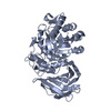
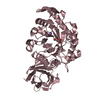
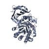
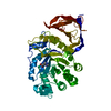
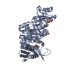
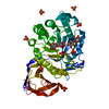
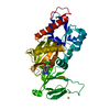
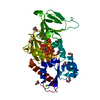
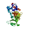
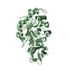
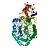
| Title | Crystal structure of Ligand bound XynC | ||||||
 Components Components | Glucuronoxylanase xynC | ||||||
 Keywords Keywords | HYDROLASE / alpha-beta barrel / (beta/alpha)8 barrel / (beta/alpha)8 + beta motif / Glucuronate coordination by XynC / Glycosidase / Polysaccharide degradation / glycosyl hydrolase | ||||||
| Function / homology |  Function and homology information Function and homology informationglucuronoarabinoxylan endo-1,4-beta-xylanase / glucuronoarabinoxylan endo-1,4-beta-xylanase activity / glucosylceramidase activity / sphingolipid metabolic process / xylan catabolic process / extracellular region / membrane Similarity search - Function | ||||||
| Biological species |  | ||||||
| Method |  X-RAY DIFFRACTION / SYNCHROTRON / MOLECULAR REPLACEMENT / molecular replacement / Resolution: 2.33 Å X-RAY DIFFRACTION / SYNCHROTRON / MOLECULAR REPLACEMENT / molecular replacement / Resolution: 2.33 Å | ||||||
 Authors Authors | St John, F.J. / Hurlbert, J.C. / Pozharski, E. | ||||||
 Citation Citation | Journal: J.Mol.Biol. / Year: 2011 Title: Ligand bound structures of a glycosyl hydrolase family 30 glucuronoxylan xylanohydrolase. Authors: St John, F.J. / Hurlbert, J.C. / Rice, J.D. / Preston, J.F. / Pozharski, E. | ||||||
| History |
|
- Structure visualization
| Structure viewer | Molecule: MolmilJmol/JSmol |
|---|
- Downloads & links
-Download
| PDBx/mmCIF format | 3kl3.cif.gz | 391.5 KB | Display | PDBx/mmCIF format |
|---|---|---|---|---|
| PDB format | pdb3kl3.ent.gz | 320.9 KB | Display | PDB format |
| PDBx/mmJSON format | 3kl3.json.gz | Tree view | PDBx/mmJSON format | |
| Others |  Other downloads Other downloads |
-Validation report
| Arichive directory | https://data.pdbj.org/pub/pdb/validation_reports/kl/3kl3ftp://data.pdbj.org/pub/pdb/validation_reports/kl/3kl3 | HTTPS FTP |
|---|
-Related structure data
| Related structure data |  3kl0SC  3kl5C S: Starting model for refinement C: citing same article ( |
|---|---|
| Similar structure data |
-Links
 PDBj
PDBj- Assembly
| Deposited unit | 
| ||||||||
|---|---|---|---|---|---|---|---|---|---|
| 1 | 
| ||||||||
| 2 | 
| ||||||||
| 3 | 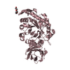
| ||||||||
| 4 | 
| ||||||||
| Unit cell |
|
-Components
-Protein , 1 types, 4 molecules ABCD
| #1: Protein | Mass: 45431.465 Da / Num. of mol.: 4 Source method: isolated from a genetically manipulated source Source: (gene. exp.) References: UniProt: Q45070, glucuronoarabinoxylan endo-1,4-beta-xylanase |
|---|
-Sugars , 2 types, 3 molecules 
| #2: Sugar |  Type: D-saccharide, alpha linking / Mass: 194.139 Da / Num. of mol.: 2 Type: D-saccharide, alpha linking / Mass: 194.139 Da / Num. of mol.: 2Source method: isolated from a genetically manipulated source Formula: C6H10O7 #4: Sugar | ChemComp-BDP / |  Type: D-saccharide, beta linking / Mass: 194.139 Da / Num. of mol.: 1 Type: D-saccharide, beta linking / Mass: 194.139 Da / Num. of mol.: 1Source method: isolated from a genetically manipulated source Formula: C6H10O7 |
|---|
-Non-polymers , 3 types, 461 molecules 


| #3: Chemical |  Type: D-peptide linking / Mass: 156.162 Da / Num. of mol.: 2 / Source method: obtained synthetically / Formula: C6H10N3O2 Type: D-peptide linking / Mass: 156.162 Da / Num. of mol.: 2 / Source method: obtained synthetically / Formula: C6H10N3O2#5: Chemical |  Mass: 194.226 Da / Num. of mol.: 2 / Source method: obtained synthetically / Formula: C8H18O5 / Comment: precipitant*YM Mass: 194.226 Da / Num. of mol.: 2 / Source method: obtained synthetically / Formula: C8H18O5 / Comment: precipitant*YM#6: Water | ChemComp-HOH / | Mass: 18.015 Da / Num. of mol.: 457 / Source method: isolated from a natural source / Formula: H2O |
|---|
-Experimental details
-Experiment
| Experiment | Method: X-RAY DIFFRACTION / Number of used crystals: 1 |
|---|
- Sample preparation
| Crystal | Density Matthews: 2.4 Å3/Da / Density % sol: 48.67 % |
|---|---|
| Crystal grow | Temperature: 294.6 K / Method: vapor diffusion, sitting drop Details: 200 mM sodium tartrate, 200 mM sodium malonate, pH 7.0, 19% PEG3350 Soaked in mother liquor/cryoprotectant/glucuronic acid solution, VAPOR DIFFUSION, SITTING DROP, temperature 294.6K |
-Data collection
| Diffraction | Mean temperature: 105 K | ||||||||||||||||||||||||||||||||||||||||||||||||||||||||||||||||||
|---|---|---|---|---|---|---|---|---|---|---|---|---|---|---|---|---|---|---|---|---|---|---|---|---|---|---|---|---|---|---|---|---|---|---|---|---|---|---|---|---|---|---|---|---|---|---|---|---|---|---|---|---|---|---|---|---|---|---|---|---|---|---|---|---|---|---|---|
| Diffraction source | Source: SYNCHROTRON / Site: SSRL  / Beamline: BL12-2 / Wavelength: 0.97946 Å / Beamline: BL12-2 / Wavelength: 0.97946 Å | ||||||||||||||||||||||||||||||||||||||||||||||||||||||||||||||||||
| Detector | Type: RAYONIX MX-325 / Detector: CCD / Date: Feb 1, 2009 | ||||||||||||||||||||||||||||||||||||||||||||||||||||||||||||||||||
| Radiation | Protocol: SINGLE WAVELENGTH / Monochromatic (M) / Laue (L): M / Scattering type: x-ray | ||||||||||||||||||||||||||||||||||||||||||||||||||||||||||||||||||
| Radiation wavelength | Wavelength: 0.97946 Å / Relative weight: 1 | ||||||||||||||||||||||||||||||||||||||||||||||||||||||||||||||||||
| Reflection | Resolution: 2.33→50 Å / Num. obs: 75511 / % possible obs: 100 % / Redundancy: 7.3 % / Rmerge(I) obs: 0.145 / Χ2: 1.079 / Net I/σ(I): 7.3 | ||||||||||||||||||||||||||||||||||||||||||||||||||||||||||||||||||
| Reflection shell |
|
-Phasing
| Phasing | Method: molecular replacement |
|---|
- Processing
| Software |
| ||||||||||||||||||||||||||||||||||||||||||||||||||||||||||||||||||||||||||||||||||||||||||||||||||||||||||||||||||||||||||||||||||||||||||||||||||||||
|---|---|---|---|---|---|---|---|---|---|---|---|---|---|---|---|---|---|---|---|---|---|---|---|---|---|---|---|---|---|---|---|---|---|---|---|---|---|---|---|---|---|---|---|---|---|---|---|---|---|---|---|---|---|---|---|---|---|---|---|---|---|---|---|---|---|---|---|---|---|---|---|---|---|---|---|---|---|---|---|---|---|---|---|---|---|---|---|---|---|---|---|---|---|---|---|---|---|---|---|---|---|---|---|---|---|---|---|---|---|---|---|---|---|---|---|---|---|---|---|---|---|---|---|---|---|---|---|---|---|---|---|---|---|---|---|---|---|---|---|---|---|---|---|---|---|---|---|---|---|---|---|
| Refinement | Method to determine structure: MOLECULAR REPLACEMENT Starting model: PDB ENTRY 3KL0 Resolution: 2.33→50 Å / Cor.coef. Fo:Fc: 0.944 / Cor.coef. Fo:Fc free: 0.917 / Occupancy max: 1 / Occupancy min: 0.3 / SU B: 7.106 / SU ML: 0.171 / Cross valid method: THROUGHOUT / σ(F): 0 / ESU R: 0.368 / ESU R Free: 0.249 / Stereochemistry target values: MAXIMUM LIKELIHOOD Details: HYDROGENS HAVE BEEN ADDED IN THE RIDING POSITIONS U VALUES : RESIDUAL ONLY
| ||||||||||||||||||||||||||||||||||||||||||||||||||||||||||||||||||||||||||||||||||||||||||||||||||||||||||||||||||||||||||||||||||||||||||||||||||||||
| Solvent computation | Ion probe radii: 0.8 Å / Shrinkage radii: 0.8 Å / VDW probe radii: 1.4 Å / Solvent model: MASK | ||||||||||||||||||||||||||||||||||||||||||||||||||||||||||||||||||||||||||||||||||||||||||||||||||||||||||||||||||||||||||||||||||||||||||||||||||||||
| Displacement parameters | Biso max: 89.7 Å2 / Biso mean: 42.939 Å2 / Biso min: 2 Å2
| ||||||||||||||||||||||||||||||||||||||||||||||||||||||||||||||||||||||||||||||||||||||||||||||||||||||||||||||||||||||||||||||||||||||||||||||||||||||
| Refinement step | Cycle: LAST / Resolution: 2.33→50 Å
| ||||||||||||||||||||||||||||||||||||||||||||||||||||||||||||||||||||||||||||||||||||||||||||||||||||||||||||||||||||||||||||||||||||||||||||||||||||||
| Refine LS restraints |
| ||||||||||||||||||||||||||||||||||||||||||||||||||||||||||||||||||||||||||||||||||||||||||||||||||||||||||||||||||||||||||||||||||||||||||||||||||||||
| LS refinement shell | Resolution: 2.33→2.391 Å / Total num. of bins used: 20
| ||||||||||||||||||||||||||||||||||||||||||||||||||||||||||||||||||||||||||||||||||||||||||||||||||||||||||||||||||||||||||||||||||||||||||||||||||||||
| Refinement TLS params. | Method: refined / Refine-ID: X-RAY DIFFRACTION
| ||||||||||||||||||||||||||||||||||||||||||||||||||||||||||||||||||||||||||||||||||||||||||||||||||||||||||||||||||||||||||||||||||||||||||||||||||||||
| Refinement TLS group |
|
