| Entry | Database: PDB / ID: 2iyf
|
|---|
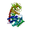
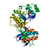
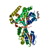
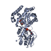
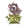
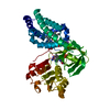
| Title | The crystal structure of macrolide glycosyltransferases: A blueprint for antibiotic engineering |
|---|
 Components Components | OLEANDOMYCIN GLYCOSYLTRANSFERASE |
|---|
 Keywords Keywords | TRANSFERASE / ANTIBIOTIC RESISTANCE / GLYCOSYLATION / GLYCOSYLTRANSFERASE / ENZYME / MACROLIDE / CARBOHYDRATE |
|---|
| Function / homology |  Function and homology information Function and homology information
UDP-glycosyltransferase activity / hexosyltransferase activity / Transferases; Glycosyltransferases; Hexosyltransferases / antibiotic biosynthetic process / response to antibioticSimilarity search - Function UDP-glycosyltransferase, MGT-like / : / Erythromycin biosynthesis protein CIII-like, central / Erythromycin biosynthesis protein CIII-like, C-terminal domain / UDP-glycosyltransferase family, conserved site / UDP-glycosyltransferases signature. / UDP-glucoronosyl and UDP-glucosyl transferase / UDP-glucuronosyl/UDP-glucosyltransferase / Glycogen Phosphorylase B; / Rossmann fold ...UDP-glycosyltransferase, MGT-like / : / Erythromycin biosynthesis protein CIII-like, central / Erythromycin biosynthesis protein CIII-like, C-terminal domain / UDP-glycosyltransferase family, conserved site / UDP-glycosyltransferases signature. / UDP-glucoronosyl and UDP-glucosyl transferase / UDP-glucuronosyl/UDP-glucosyltransferase / Glycogen Phosphorylase B; / Rossmann fold / 3-Layer(aba) Sandwich / Alpha BetaSimilarity search - Domain/homology |
|---|
| Biological species |  STREPTOMYCES ANTIBIOTICUS (bacteria) STREPTOMYCES ANTIBIOTICUS (bacteria) |
|---|
| Method |  X-RAY DIFFRACTION / SYNCHROTRON / MOLECULAR REPLACEMENT / Resolution: 1.7 Å X-RAY DIFFRACTION / SYNCHROTRON / MOLECULAR REPLACEMENT / Resolution: 1.7 Å |
|---|
 Authors Authors | Bolam, D.N. / Roberts, S.M. / Proctor, M.R. / Turkenburg, J.P. / Dodson, E.J. / Martinez-Fleites, C. / Yang, M. / Davis, B.G. / Davies, G.J. / Gilbert, H.J. |
|---|
 Citation Citation | Journal: Proc.Natl.Acad.Sci.USA / Year: 2007
Title: The Crystal Structure of Two Macrolide Glycosyltransferases Provides a Blueprint for Host Cell Antibiotic Immunity.
Authors: Bolam, D.N. / Roberts, S.M. / Proctor, M.R. / Turkenburg, J.P. / Dodson, E.J. / Martinez-Fleites, C. / Yang, M. / Davis, B.G. / Davies, G.J. / Gilbert, H.J. |
|---|
| History | | Deposition | Jul 17, 2006 | Deposition site: PDBE / Processing site: PDBE |
|---|
| Revision 1.0 | Mar 27, 2007 | Provider: repository / Type: Initial release |
|---|
| Revision 1.1 | Aug 7, 2013 | Group: Database references / Derived calculations ...Database references / Derived calculations / Other / Source and taxonomy / Structure summary / Version format compliance |
|---|
| Revision 1.2 | May 8, 2024 | Group: Data collection / Database references ...Data collection / Database references / Derived calculations / Other
Category: chem_comp_atom / chem_comp_bond ...chem_comp_atom / chem_comp_bond / database_2 / pdbx_database_status / pdbx_struct_conn_angle / struct_conn
Item: _database_2.pdbx_DOI / _database_2.pdbx_database_accession ..._database_2.pdbx_DOI / _database_2.pdbx_database_accession / _pdbx_database_status.status_code_sf / _pdbx_struct_conn_angle.ptnr1_auth_comp_id / _pdbx_struct_conn_angle.ptnr1_auth_seq_id / _pdbx_struct_conn_angle.ptnr1_label_asym_id / _pdbx_struct_conn_angle.ptnr1_label_comp_id / _pdbx_struct_conn_angle.ptnr1_label_seq_id / _pdbx_struct_conn_angle.ptnr3_auth_comp_id / _pdbx_struct_conn_angle.ptnr3_auth_seq_id / _pdbx_struct_conn_angle.ptnr3_label_asym_id / _pdbx_struct_conn_angle.ptnr3_label_comp_id / _pdbx_struct_conn_angle.ptnr3_label_seq_id / _pdbx_struct_conn_angle.value / _struct_conn.pdbx_dist_value / _struct_conn.ptnr1_auth_comp_id / _struct_conn.ptnr1_auth_seq_id / _struct_conn.ptnr1_label_asym_id / _struct_conn.ptnr1_label_atom_id / _struct_conn.ptnr1_label_comp_id / _struct_conn.ptnr1_label_seq_id / _struct_conn.ptnr2_auth_comp_id / _struct_conn.ptnr2_auth_seq_id / _struct_conn.ptnr2_label_asym_id / _struct_conn.ptnr2_label_atom_id / _struct_conn.ptnr2_label_comp_id / _struct_conn.ptnr2_label_seq_id |
|---|
|
|---|
 Movie
Movie Controller

 Yorodumi
 Open data
 Basic information
 Structure visualization
 Downloads & links
 Other downloads
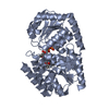


 PDBj
PDBj


 Assembly


 Mass: 733.927 Da / Num. of mol.: 2 / Source method: obtained synthetically / Formula: C37H67NO13 / Comment: antibiotic*YM
 Type: RNA linking / Mass: 404.161 Da / Num. of mol.: 2 / Source method: obtained synthetically / Formula: C9H14N2O12P2 / Comment: UDP*YM
 Mass: 24.305 Da / Num. of mol.: 1 / Source method: obtained synthetically / Formula: Mg
 Mass: 18.015 Da / Num. of mol.: 604 / Source method: isolated from a natural source / Formula: H2O
 Sample preparation
 / Beamline: ID14-3 / Wavelength: 0.933
  Processing