 Movie
Movie Controller

[English] 日本語
 Yorodumi
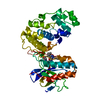
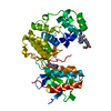
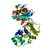
- PDB-3o8t: Conformational plasticity of p38 MAP kinase DFG-motif mutants in ... -
+ Open data

- Basic information
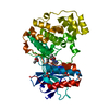
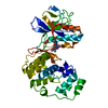
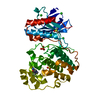
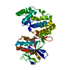
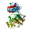
| Entry | Database: PDB / ID: 3o8t | ||||||
|---|---|---|---|---|---|---|---|
| Title | Conformational plasticity of p38 MAP kinase DFG-motif mutants in response to inhibitor binding | ||||||
 Components Components | Mitogen-activated protein kinase 14 | ||||||
 Keywords Keywords | TRANSFERASE | ||||||
| Function / homology |  Function and homology information Function and homology informationpositive regulation of cyclase activity / Activation of PPARGC1A (PGC-1alpha) by phosphorylation / regulation of synaptic membrane adhesion / stress-induced premature senescence / stress-activated protein kinase signaling cascade / CD163 mediating an anti-inflammatory response / 3'-UTR-mediated mRNA stabilization / : / cell surface receptor protein serine/threonine kinase signaling pathway / positive regulation of myoblast fusion ...positive regulation of cyclase activity / Activation of PPARGC1A (PGC-1alpha) by phosphorylation / regulation of synaptic membrane adhesion / stress-induced premature senescence / stress-activated protein kinase signaling cascade / CD163 mediating an anti-inflammatory response / 3'-UTR-mediated mRNA stabilization / : / cell surface receptor protein serine/threonine kinase signaling pathway / positive regulation of myoblast fusion / KSRP (KHSRP) binds and destabilizes mRNA / cellular response to UV-B / positive regulation of muscle cell differentiation / mitogen-activated protein kinase p38 binding / cartilage condensation / Myogenesis / Platelet sensitization by LDL / NFAT protein binding / positive regulation of myotube differentiation / regulation of cytokine production involved in inflammatory response / Activation of the AP-1 family of transcription factors / cellular response to lipoteichoic acid / ERK/MAPK targets / p38MAPK cascade / response to dietary excess / response to muramyl dipeptide / fatty acid oxidation / MAP kinase kinase activity / Regulation of MITF-M-dependent genes involved in pigmentation / chondrocyte differentiation / MAP kinase activity / regulation of ossification / cellular response to vascular endothelial growth factor stimulus / mitogen-activated protein kinase / vascular endothelial growth factor receptor signaling pathway / RHO GTPases Activate NADPH Oxidases / positive regulation of myoblast differentiation / negative regulation of hippo signaling / positive regulation of cardiac muscle cell proliferation / stress-activated MAPK cascade / skeletal muscle tissue development / positive regulation of interleukin-12 production / positive regulation of brown fat cell differentiation / striated muscle cell differentiation / response to muscle stretch / signal transduction in response to DNA damage / p38MAPK events / osteoclast differentiation / DNA damage checkpoint signaling / positive regulation of erythrocyte differentiation / lipopolysaccharide-mediated signaling pathway / placenta development / stem cell differentiation / positive regulation of D-glucose import across plasma membrane / tumor necrosis factor-mediated signaling pathway / cellular response to ionizing radiation / activated TAK1 mediates p38 MAPK activation / negative regulation of inflammatory response to antigenic stimulus / negative regulation of canonical Wnt signaling pathway / positive regulation of protein import into nucleus / platelet activation / response to insulin / cellular response to virus / NOD1/2 Signaling Pathway / bone development / glucose metabolic process / VEGFA-VEGFR2 Pathway / cell morphogenesis / positive regulation of reactive oxygen species metabolic process / cellular senescence / chemotaxis / osteoblast differentiation / spindle pole / MAPK cascade / ADP signalling through P2Y purinoceptor 1 / cellular response to lipopolysaccharide / transcription by RNA polymerase II / angiogenesis / secretory granule lumen / protein phosphatase binding / Oxidative Stress Induced Senescence / Regulation of TP53 Activity through Phosphorylation / ficolin-1-rich granule lumen / cell surface receptor signaling pathway / nuclear speck / intracellular signal transduction / protein serine kinase activity / protein serine/threonine kinase activity / apoptotic process / positive regulation of gene expression / Neutrophil degranulation / regulation of transcription by RNA polymerase II / glutamatergic synapse / enzyme binding / signal transduction / positive regulation of transcription by RNA polymerase II / mitochondrion / extracellular region / nucleoplasm / ATP binding Similarity search - Function | ||||||
| Biological species |  Homo sapiens (human) Homo sapiens (human) | ||||||
| Method |  X-RAY DIFFRACTION / SYNCHROTRON / MOLECULAR REPLACEMENT / Resolution: 2 Å X-RAY DIFFRACTION / SYNCHROTRON / MOLECULAR REPLACEMENT / Resolution: 2 Å | ||||||
 Authors Authors | Namboodiri, H.V. / Karpusas, M. / Bukhtiyarova, M. / Springman, E.B. | ||||||
 Citation Citation | Journal: To be Published Title: Conformational plasticity of p38 MAP kinase DFG motif mutants in response to inhibitor binding Authors: Namboodiri, H.V. / Springman, E.B. / Karpusas, M. / Bukhtiyarova, M. / Ramcharan, J. | ||||||
| History |
|
- Structure visualization
| Structure viewer | Molecule: MolmilJmol/JSmol |
|---|
- Downloads & links
-Download
| PDBx/mmCIF format | 3o8t.cif.gz | 86 KB | Display | PDBx/mmCIF format |
|---|---|---|---|---|
| PDB format | pdb3o8t.ent.gz | 62.8 KB | Display | PDB format |
| PDBx/mmJSON format | 3o8t.json.gz | Tree view | PDBx/mmJSON format | |
| Others |  Other downloads Other downloads |
-Validation report
| Arichive directory | https://data.pdbj.org/pub/pdb/validation_reports/o8/3o8tftp://data.pdbj.org/pub/pdb/validation_reports/o8/3o8t | HTTPS FTP |
|---|
-Related structure data
| Related structure data |  3mpaC  3o8pC  3o8uC  3obgC  3objC  3oc1C 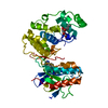 1zyjS C: citing same article ( S: Starting model for refinement |
|---|---|
| Similar structure data |


-Links
 PDBj
PDBj


- Assembly
| Deposited unit | 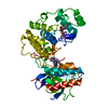
| ||||||||
|---|---|---|---|---|---|---|---|---|---|
| 1 |
| ||||||||
| Unit cell |
|
-Components
| #1: Protein | Mass: 41267.102 Da / Num. of mol.: 1 / Fragment: kinase domain / Mutation: F169A Source method: isolated from a genetically manipulated source Source: (gene. exp.) Homo sapiens (human) / Gene: CSBP, CSBP1, CSBP2, CSPB1, MAPK14, MXI2References: UniProt: Q16539, mitogen-activated protein kinase |
|---|---|
| #2: Chemical | ChemComp-BMU / 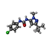  Mass: 306.791 Da / Num. of mol.: 1 / Source method: obtained synthetically / Formula: C15H19ClN4O Mass: 306.791 Da / Num. of mol.: 1 / Source method: obtained synthetically / Formula: C15H19ClN4O |
| #3: Sugar | ChemComp-BOG /   Type: D-saccharide / Mass: 292.369 Da / Num. of mol.: 1 Type: D-saccharide / Mass: 292.369 Da / Num. of mol.: 1Source method: isolated from a genetically manipulated source Formula: C14H28O6 / Comment: detergent*YM |
| #4: Water | ChemComp-HOH /  Mass: 18.015 Da / Num. of mol.: 162 / Source method: isolated from a natural source / Formula: H2O Mass: 18.015 Da / Num. of mol.: 162 / Source method: isolated from a natural source / Formula: H2O |
-Experimental details
-Experiment
| Experiment | Method: X-RAY DIFFRACTION / Number of used crystals: 1 |
|---|
- Sample preparation
| Crystal | Density Matthews: 2.22 Å3/Da / Density % sol: 44.61 % |
|---|---|
| Crystal grow | Temperature: 295 K / Method: vapor diffusion, sitting drop / pH: 6 Details: 10-20% PEG4000, 0.1M Cacodylic acid, 50 mM n-octyl-beta-D-glucoside, pH 6.0, VAPOR DIFFUSION, SITTING DROP, temperature 295K |
-Data collection
| Diffraction | Mean temperature: 93 K |
|---|---|
| Diffraction source | Source: SYNCHROTRON / Site: NSLS  / Beamline: X4A / Wavelength: 0.98 Å / Beamline: X4A / Wavelength: 0.98 Å |
| Detector | Type: ADSC QUANTUM 4 / Detector: CCD / Details: Mirrors |
| Radiation | Protocol: SINGLE WAVELENGTH / Monochromatic (M) / Laue (L): M / Scattering type: x-ray |
| Radiation wavelength | Wavelength: 0.98 Å / Relative weight: 1 |
| Reflection | Resolution: 2→37.84 Å / Num. obs: 25513 / % possible obs: 90.8 % / Observed criterion σ(F): 1 / Observed criterion σ(I): 1 / Redundancy: 4.5 % / Rmerge(I) obs: 0.05 / Rsym value: 0.046 / Net I/σ(I): 30 |
| Reflection shell | Resolution: 2→2.15 Å / Redundancy: 4.4 % / Mean I/σ(I) obs: 5 / Num. unique all: 4677 / % possible all: 93.4 |
- Processing
| Software |
| ||||||||||||||||||||||||||||||||||||||||||||||||||||||||||||||||||||||||||||||||||||||||||
|---|---|---|---|---|---|---|---|---|---|---|---|---|---|---|---|---|---|---|---|---|---|---|---|---|---|---|---|---|---|---|---|---|---|---|---|---|---|---|---|---|---|---|---|---|---|---|---|---|---|---|---|---|---|---|---|---|---|---|---|---|---|---|---|---|---|---|---|---|---|---|---|---|---|---|---|---|---|---|---|---|---|---|---|---|---|---|---|---|---|---|---|
| Refinement | Method to determine structure: MOLECULAR REPLACEMENT Starting model: PDB entry 1ZYJ Resolution: 2→37.84 Å / Cor.coef. Fo:Fc: 0.938 / Cor.coef. Fo:Fc free: 0.899 / SU B: 5.534 / SU ML: 0.155 / Cross valid method: THROUGHOUT / ESU R Free: 0.22 / Stereochemistry target values: MAXIMUM LIKELIHOOD
| ||||||||||||||||||||||||||||||||||||||||||||||||||||||||||||||||||||||||||||||||||||||||||
| Solvent computation | Ion probe radii: 0.8 Å / Shrinkage radii: 0.8 Å / VDW probe radii: 1.2 Å / Solvent model: MASK | ||||||||||||||||||||||||||||||||||||||||||||||||||||||||||||||||||||||||||||||||||||||||||
| Displacement parameters | Biso mean: 35.497 Å2
| ||||||||||||||||||||||||||||||||||||||||||||||||||||||||||||||||||||||||||||||||||||||||||
| Refinement step | Cycle: LAST / Resolution: 2→37.84 Å
| ||||||||||||||||||||||||||||||||||||||||||||||||||||||||||||||||||||||||||||||||||||||||||
| Refine LS restraints |
| ||||||||||||||||||||||||||||||||||||||||||||||||||||||||||||||||||||||||||||||||||||||||||
| LS refinement shell | Resolution: 2→2.052 Å / Total num. of bins used: 20
|
