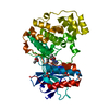
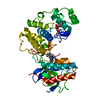
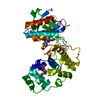
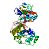
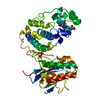
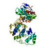
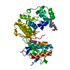
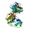
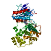
Entry Database : PDB / ID : 2puuTitle Crystal structure of p38 complex with 1-(5-tert-Butyl-2-p-tolyl-2H-pyrazol-3-yl)-3-[4-(6-morpholin-4-ylmethyl-pyridin-3-yl)naphthalen-1-yl]urea Mitogen-activated protein kinase 14 Keywords / / / Function / homology Function Domain/homology Component
/ / / / / / / / / / / / / / / / / / / / / / / / / / / / / / / / / / / / / / / / / / / / / / / / / / / / / / / / / / / / / / / / / / / / / / / / / / / / / / / / / / / / / / / / / / / / / / / / / / / / / / / / / / / / / / / / / / / / / / / / / / / / / / / / / / Biological species Mus musculus (house mouse)Method / / Resolution : 2.5 Å Authors Qian, K.C. Journal : To be Published Title : A new class of p38 MAP kinase inhibitors based on the pyrazole-naphthyl urea scaffoldAuthors : Qian, K.C. History Deposition May 9, 2007 Deposition site / Processing site Revision 1.0 May 20, 2008 Provider / Type Revision 1.1 Jul 13, 2011 Group Revision 1.2 Apr 4, 2018 Group / Category / Item Revision 1.3 Oct 20, 2021 Group / Derived calculations / Category / struct_ref_seq_dif / struct_siteItem _database_2.pdbx_DOI / _database_2.pdbx_database_accession ... _database_2.pdbx_DOI / _database_2.pdbx_database_accession / _struct_ref_seq_dif.details / _struct_site.pdbx_auth_asym_id / _struct_site.pdbx_auth_comp_id / _struct_site.pdbx_auth_seq_id Revision 1.4 Feb 21, 2024 Group / Category / chem_comp_bond
Show all Show less
 Movie
Movie Controller

 Yorodumi
 Open data
 Basic information
 Components
 Keywords
 Function and homology information
 X-RAY DIFFRACTION /
  Authors
 Citation
 Structure visualization
 Downloads & links
 Other downloads


 PDBj
PDBj


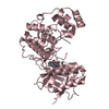
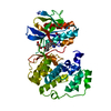
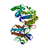
 Assembly
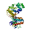

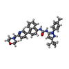
 Mass: 575.723 Da / Num. of mol.: 1 / Source method: obtained synthetically / Formula: C35H39N6O2
 Mass: 18.015 Da / Num. of mol.: 31 / Source method: isolated from a natural source / Formula: H2O
 Sample preparation
 Processing