 Movie
Movie Controller

[English] 日本語
 Yorodumi
- PDB-1btj: HUMAN SERUM TRANSFERRIN, RECOMBINANT N-TERMINAL LOBE, APO FORM, C... -
+ Open data

- Basic information
| Entry | Database: PDB / ID: 1btj | ||||||
|---|---|---|---|---|---|---|---|
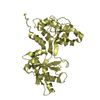
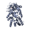
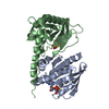
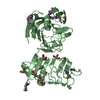
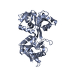
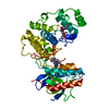
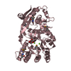
| Title | HUMAN SERUM TRANSFERRIN, RECOMBINANT N-TERMINAL LOBE, APO FORM, CRYSTAL FORM 2 | ||||||
 Components Components | PROTEIN (SERUM TRANSFERRIN) | ||||||
 Keywords Keywords | METAL TRANSPORT / IRON TRANSPORT / GLYCOPROTEIN / TRANSFERRIN / N-LOBE / IRON-FREE FORM / CONFORMATIONAL CHANGE | ||||||
| Function / homology |  Function and homology information Function and homology informationiron chaperone activity / transferrin receptor binding / Transferrin endocytosis and recycling / basal part of cell / endocytic vesicle / clathrin-coated pit / ferric iron binding / osteoclast differentiation / basal plasma membrane / Post-translational protein phosphorylation ...iron chaperone activity / transferrin receptor binding / Transferrin endocytosis and recycling / basal part of cell / endocytic vesicle / clathrin-coated pit / ferric iron binding / osteoclast differentiation / basal plasma membrane / Post-translational protein phosphorylation / iron ion transport / clathrin-coated endocytic vesicle membrane / HFE-transferrin receptor complex / cellular response to iron ion / regulation of protein stability / ferrous iron binding / Iron uptake and transport / positive regulation of receptor-mediated endocytosis / multicellular organismal-level iron ion homeostasis / recycling endosome / Regulation of Insulin-like Growth Factor (IGF) transport and uptake by Insulin-like Growth Factor Binding Proteins (IGFBPs) / Platelet degranulation / late endosome / Cargo recognition for clathrin-mediated endocytosis / positive regulation of proteasomal ubiquitin-dependent protein catabolic process / antibacterial humoral response / Clathrin-mediated endocytosis / cytoplasmic vesicle / secretory granule lumen / blood microparticle / vesicle / intracellular iron ion homeostasis / transmembrane transporter binding / early endosome / cell surface receptor signaling pathway / endosome membrane / apical plasma membrane / endoplasmic reticulum lumen / perinuclear region of cytoplasm / enzyme binding / cell surface / : / extracellular exosome / extracellular region / plasma membrane Similarity search - Function | ||||||
| Biological species |  Homo sapiens (human) Homo sapiens (human) | ||||||
| Method |  X-RAY DIFFRACTION / MOLECULAR REPLACEMENT / Resolution: 3.2 Å X-RAY DIFFRACTION / MOLECULAR REPLACEMENT / Resolution: 3.2 Å | ||||||
 Authors Authors | Jeffrey, P.D. / Bewley, M.C. / Macgillivray, R.T.A. / Mason, A.B. / Woodworth, R.C. / Baker, E.N. | ||||||
 Citation Citation | Journal: Biochemistry / Year: 1998 Title: Ligand-induced conformational change in transferrins: crystal structure of the open form of the N-terminal half-molecule of human transferrin. Authors: Jeffrey, P.D. / Bewley, M.C. / MacGillivray, R.T. / Mason, A.B. / Woodworth, R.C. / Baker, E.N. | ||||||
| History |
|
- Structure visualization
| Structure viewer | Molecule: MolmilJmol/JSmol |
|---|
- Downloads & links
-Download
| PDBx/mmCIF format | 1btj.cif.gz | 122 KB | Display | PDBx/mmCIF format |
|---|---|---|---|---|
| PDB format | pdb1btj.ent.gz | 97.5 KB | Display | PDB format |
| PDBx/mmJSON format | 1btj.json.gz | Tree view | PDBx/mmJSON format | |
| Others |  Other downloads Other downloads |
-Validation report
| Arichive directory | https://data.pdbj.org/pub/pdb/validation_reports/bt/1btjftp://data.pdbj.org/pub/pdb/validation_reports/bt/1btj | HTTPS FTP |
|---|
-Related structure data


-Links
 PDBj
PDBj


- Assembly
| Deposited unit | 
| ||||||||
|---|---|---|---|---|---|---|---|---|---|
| 1 | 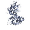
| ||||||||
| 2 | 
| ||||||||
| Unit cell |
|
-Components
| #1: Protein | Mass: 37215.270 Da / Num. of mol.: 2 / Fragment: N-TERMINAL LOBE Source method: isolated from a genetically manipulated source Source: (gene. exp.) Homo sapiens (human) / Production host:  Cricetinae (hamsters) / References: UniProt: P02787 Cricetinae (hamsters) / References: UniProt: P02787Has protein modification | Y | |
|---|
-Experimental details
-Experiment
| Experiment | Method: X-RAY DIFFRACTION / Number of used crystals: 1 |
|---|
- Sample preparation
| Crystal | Density Matthews: 2.27 Å3/Da / Density % sol: 46 % | ||||||||||||||||||||||||||||||||||||
|---|---|---|---|---|---|---|---|---|---|---|---|---|---|---|---|---|---|---|---|---|---|---|---|---|---|---|---|---|---|---|---|---|---|---|---|---|---|
| Crystal grow | Method: vapor diffusion, sitting drop / pH: 5.3 Details: PROTEIN DROP: 20 MG/ML PROTEIN, 20MM NAHCO3, 50 MM KCL. RESERVOIR: 50 MM POTASSIUM ACETATE, PH 5.3, 35% MPD., VAPOR DIFFUSION, SITTING DROP | ||||||||||||||||||||||||||||||||||||
| Crystal | *PLUS Density % sol: 46 % | ||||||||||||||||||||||||||||||||||||
| Crystal grow | *PLUS Temperature: 4 ℃ | ||||||||||||||||||||||||||||||||||||
| Components of the solutions | *PLUS
|
-Data collection
| Diffraction | Mean temperature: 113 K |
|---|---|
| Diffraction source | Source: ROTATING ANODE / Type: RIGAKU RU200 / Wavelength: 1.5418 |
| Detector | Type: RIGAKU RAXIS IIC / Detector: IMAGE PLATE / Date: May 15, 1996 |
| Radiation | Monochromator: GRAPHITE CRYSTAL / Protocol: SINGLE WAVELENGTH / Monochromatic (M) / Laue (L): M / Scattering type: x-ray |
| Radiation wavelength | Wavelength: 1.5418 Å / Relative weight: 1 |
| Reflection | Resolution: 3.2→40 Å / Num. obs: 11269 / % possible obs: 99.7 % / Observed criterion σ(I): 0 / Redundancy: 3.3 % / Rmerge(I) obs: 0.111 / Net I/σ(I): 5.9 |
| Reflection shell | Resolution: 3.2→3.5 Å / Redundancy: 3.3 % / Rmerge(I) obs: 0.257 / Mean I/σ(I) obs: 2.6 / % possible all: 99.9 |
| Reflection shell | *PLUS % possible obs: 99.9 % |
- Processing
| Software |
| ||||||||||||||||||||||||||||||||||||||||||||||||||||||||||||
|---|---|---|---|---|---|---|---|---|---|---|---|---|---|---|---|---|---|---|---|---|---|---|---|---|---|---|---|---|---|---|---|---|---|---|---|---|---|---|---|---|---|---|---|---|---|---|---|---|---|---|---|---|---|---|---|---|---|---|---|---|---|
| Refinement | Method to determine structure: MOLECULAR REPLACEMENT Starting model: HUMAN TRANSFERRIN N-LOBE, HOLO FORM, INDIVIDUAL DOMAINS Resolution: 3.2→6 Å / Cross valid method: R-FREE / σ(F): 0
| ||||||||||||||||||||||||||||||||||||||||||||||||||||||||||||
| Refine analyze | Luzzati d res low obs: 6 Å | ||||||||||||||||||||||||||||||||||||||||||||||||||||||||||||
| Refinement step | Cycle: LAST / Resolution: 3.2→6 Å
| ||||||||||||||||||||||||||||||||||||||||||||||||||||||||||||
| Refine LS restraints |
| ||||||||||||||||||||||||||||||||||||||||||||||||||||||||||||
| Refine LS restraints NCS | NCS model details: RESTRAINTS | ||||||||||||||||||||||||||||||||||||||||||||||||||||||||||||
| Software | *PLUS Name: X-PLOR / Version: 3.1 / Classification: refinement | ||||||||||||||||||||||||||||||||||||||||||||||||||||||||||||
| Refinement | *PLUS Highest resolution: 3.2 Å / Lowest resolution: 6 Å / σ(F): 0 / % reflection Rfree: 10 % | ||||||||||||||||||||||||||||||||||||||||||||||||||||||||||||
| Solvent computation | *PLUS | ||||||||||||||||||||||||||||||||||||||||||||||||||||||||||||
| Displacement parameters | *PLUS | ||||||||||||||||||||||||||||||||||||||||||||||||||||||||||||
| Refine LS restraints | *PLUS Type: x_angle_deg / Dev ideal: 1.9 |
