Protocol: SINGLE WAVELENGTH / Monochromatic (M) / Laue (L): M / Scattering type: x-ray
Radiation wavelength
Wavelength: 0.97945 Å / Relative weight: 1
Reflection
Resolution: 1.7→51.71 Å / Num. obs: 81807 / % possible obs: 96.1 % / Observed criterion σ(I): 2 / Redundancy: 4.2 % / Rmerge(I) obs: 0.08 / Net I/σ(I): 13.7
Reflection shell
Resolution: 1.7→1.79 Å / Redundancy: 3.8 % / Rmerge(I) obs: 0.26 / Mean I/σ(I) obs: 4 / % possible all: 80.1
-
Processing
Software
Name
Version
Classification
REFMAC
5.2.0019
refinement
MOSFLM
datareduction
SCALA
datascaling
SHELX
phasing
Refinement
Method to determine structure: SAD / Resolution: 1.7→74.54 Å / Cor.coef. Fo:Fc: 0.957 / Cor.coef. Fo:Fc free: 0.943 / SU B: 1.845 / SU ML: 0.062 / Cross valid method: THROUGHOUT / ESU R: 0.106 / ESU R Free: 0.102 / Stereochemistry target values: MAXIMUM LIKELIHOOD / Details: HYDROGENS HAVE BEEN ADDED IN THE RIDING POSITIONS.
Rfactor
Num. reflection
% reflection
Selection details
Rfree
0.198
4084
5 %
RANDOM
Rwork
0.167
-
-
-
obs
0.168
77514
95.8 %
-
Solvent computation
Ion probe radii: 0.8 Å / Shrinkage radii: 0.8 Å / VDW probe radii: 1.4 Å / Solvent model: MASK
 Movie
Movie Controller

 Yorodumi
 Open data
 Basic information
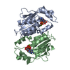
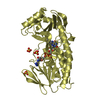
 Components
 Keywords
 Function and homology information
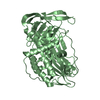
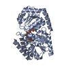
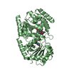
 STREPTOMYCES ANTIBIOTICUS (bacteria)
 X-RAY DIFFRACTION /
  Authors
 Citation
 Structure visualization
 Downloads & links
 Other downloads
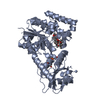


 PDBj
PDBj


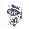
 Assembly

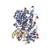
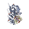
 Type: RNA linking / Mass: 404.161 Da / Num. of mol.: 2 / Source method: obtained synthetically / Formula: C9H14N2O12P2 / Comment: UDP*YM
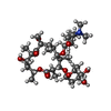
 Mass: 687.858 Da / Num. of mol.: 2 / Source method: obtained synthetically / Formula: C35H61NO12
 Mass: 18.015 Da / Num. of mol.: 642 / Source method: isolated from a natural source / Formula: H2O
 Sample preparation
 / Beamline: ID23-1 / Wavelength: 0.97945
  Processing