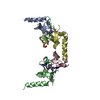
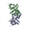
Entry Database : PDB / ID : 2ix7Title Structure of apo-calmodulin bound to unconventional myosin V Keywords / / / / / / / / / / / / / / / / / / / Function / homology Function Domain/homology Component
/ / / / / / / / / / / / / / / / / / / / / / / / / / / / / / / / / / / / / / / / / / / / / / / / / / / / / / / / / / / / / / / / / / / / / / / / / / / / / / / / / / / / / / / / / / / / / / / / / / / / / / / / / / / / / / / / / / / / / / / / / / / / / / / / / / / / / / / / / / / / / / / / / / / / / / / Biological species MUS MUSCULUS (house mouse)Method / / / Resolution : 2.5 Å Authors Houdusse, A. / Gaucher, J.F. / Mui, S. / Krementsova, E. / Trybus, K.M. / Cohen, C. Journal : Proc.Natl.Acad.Sci.USA / Year : 2006Title : Crystal Structure of Apo-Calmodulin Bound to the First Two Iq Motifs of Myosin V Reveals Essential Recognition Features.Authors : Houdusse, A. / Gaucher, J.F. / Krementsova, E. / Mui, S. / Trybus, K.M. / Cohen, C. History Deposition Jul 7, 2006 Deposition site / Processing site Revision 1.0 Dec 13, 2006 Provider / Type Revision 1.1 Dec 4, 2013 Group Data collection / Derived calculations ... Data collection / Derived calculations / Non-polymer description / Other / Refinement description / Source and taxonomy / Version format compliance Revision 1.2 Feb 27, 2019 Group Data collection / Derived calculations ... Data collection / Derived calculations / Experimental preparation / Other Category exptl_crystal_grow / pdbx_database_proc ... exptl_crystal_grow / pdbx_database_proc / pdbx_database_status / struct_biol / struct_conn Item _exptl_crystal_grow.method / _exptl_crystal_grow.temp ... _exptl_crystal_grow.method / _exptl_crystal_grow.temp / _pdbx_database_status.recvd_author_approval / _struct_conn.pdbx_leaving_atom_flag Revision 1.3 Nov 8, 2023 Group Data collection / Database references ... Data collection / Database references / Derived calculations / Other Category chem_comp_atom / chem_comp_bond ... chem_comp_atom / chem_comp_bond / database_2 / pdbx_database_status / struct_conn / struct_conn_type / struct_site Item _database_2.pdbx_DOI / _database_2.pdbx_database_accession ... _database_2.pdbx_DOI / _database_2.pdbx_database_accession / _pdbx_database_status.status_code_sf / _struct_conn.conn_type_id / _struct_conn.id / _struct_conn_type.id / _struct_site.pdbx_auth_asym_id / _struct_site.pdbx_auth_comp_id / _struct_site.pdbx_auth_seq_id Revision 1.4 Dec 13, 2023 Group / Category Revision 1.5 Oct 23, 2024 Group / Category / pdbx_modification_feature
Show all Show less
 Movie
Movie Controller


 Open data
 Basic information
 Components
 Keywords
 Function and homology information
 X-RAY DIFFRACTION /
  Authors
 Citation
 Structure visualization
 Downloads & links
 Other downloads


 PDBj
PDBj


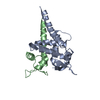
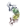
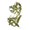
 Assembly


 Mass: 96.063 Da / Num. of mol.: 5 / Source method: obtained synthetically / Formula: SO4
 Type: L-peptide linking / Mass: 121.158 Da / Num. of mol.: 1 / Source method: obtained synthetically / Formula: C3H7NO2S
 Mass: 18.015 Da / Num. of mol.: 144 / Source method: isolated from a natural source / Formula: H2O
 Sample preparation
 / Beamline: A1 / Wavelength: 0.9134
  Processing