 Movie
Movie Controller

[English] 日本語
 Yorodumi
- PDB-6hn7: Hijacking the Hijackers: Escherichia coli Pathogenicity Islands R... -
+ Open data

- Basic information
| Entry | Database: PDB / ID: 6hn7 | ||||||
|---|---|---|---|---|---|---|---|
| Title | Hijacking the Hijackers: Escherichia coli Pathogenicity Islands Redirect Helper Phage Packaging for Their Own Benefit. | ||||||
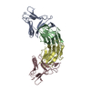
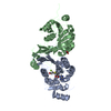
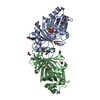
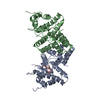
 Components Components |
| ||||||
 Keywords Keywords | DNA BINDING PROTEIN / Redirecting packaging protein / Hetero-dimer / phage interference / Protein complex | ||||||
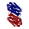
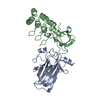
| Function / homology |  Function and homology information Function and homology informationviral terminase, small subunit / sequence-specific DNA binding, bending / viral DNA genome packaging / Hydrolases; Acting on acid anhydrides; Acting on acid anhydrides to facilitate cellular and subcellular movement / host cell cytoplasm / ATP hydrolysis activity / ATP binding Similarity search - Function | ||||||
| Biological species |   Escherichia virus Lambda Escherichia virus Lambda | ||||||
| Method |  X-RAY DIFFRACTION / SYNCHROTRON / MOLECULAR REPLACEMENT / Resolution: 3 Å X-RAY DIFFRACTION / SYNCHROTRON / MOLECULAR REPLACEMENT / Resolution: 3 Å | ||||||
 Authors Authors | Penades, J.R. / Bacarizo, J. / Marina, A. / Alqasmi, M. / Fillol-Salom, A. / Roszak, A.W. / Ciges-Tomas, J.R. | ||||||
| Funding support |  United Kingdom, 1items United Kingdom, 1items
| ||||||
 Citation Citation | Journal: Mol.Cell / Year: 2019 Title: Hijacking the Hijackers: Escherichia coli Pathogenicity Islands Redirect Helper Phage Packaging for Their Own Benefit. Authors: Fillol-Salom, A. / Bacarizo, J. / Alqasmi, M. / Ciges-Tomas, J.R. / Martinez-Rubio, R. / Roszak, A.W. / Cogdell, R.J. / Chen, J. / Marina, A. / Penades, J.R. | ||||||
| History |
|
- Structure visualization
| Structure viewer | Molecule: MolmilJmol/JSmol |
|---|
- Downloads & links
-Download
| PDBx/mmCIF format | 6hn7.cif.gz | 56.5 KB | Display | PDBx/mmCIF format |
|---|---|---|---|---|
| PDB format | pdb6hn7.ent.gz | 38.2 KB | Display | PDB format |
| PDBx/mmJSON format | 6hn7.json.gz | Tree view | PDBx/mmJSON format | |
| Others |  Other downloads Other downloads |
-Validation report
| Arichive directory | https://data.pdbj.org/pub/pdb/validation_reports/hn/6hn7ftp://data.pdbj.org/pub/pdb/validation_reports/hn/6hn7 | HTTPS FTP |
|---|
-Related structure data
| Related structure data | 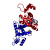 6hlkSC 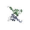 1j9iS S: Starting model for refinement C: citing same article ( |
|---|---|
| Similar structure data |


-Links
 PDBj
PDBj

- Assembly
| Deposited unit | 
| ||||||||||
|---|---|---|---|---|---|---|---|---|---|---|---|
| 1 | 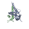
| ||||||||||
| Unit cell |
|
-Components
| #1: Protein | Mass: 17828.396 Da / Num. of mol.: 1 Source method: isolated from a genetically manipulated source Source: (gene. exp.) Production host: References: UniProt: A0A5H1ZR32*PLUS |
|---|---|
| #2: Protein | Mass: 11472.711 Da / Num. of mol.: 1 Source method: isolated from a genetically manipulated source Details: All alpha proteins, Putative DNA-binding domain, Hetero-dimer complex, Terminase gpNU1 subunit domain, Bacteriophage lambda The first three Ala residues at the beginning of the sequence from ...Details: All alpha proteins, Putative DNA-binding domain, Hetero-dimer complex, Terminase gpNU1 subunit domain, Bacteriophage lambda The first three Ala residues at the beginning of the sequence from coordinates come from the purification tag. Source: (gene. exp.) Escherichia virus Lambda / Gene: Nu1, lambdap01Production host: References: UniProt: P03707 |
| #3: Water | ChemComp-HOH /  Mass: 18.015 Da / Num. of mol.: 20 / Source method: isolated from a natural source / Formula: H2O Mass: 18.015 Da / Num. of mol.: 20 / Source method: isolated from a natural source / Formula: H2O |
-Experimental details
-Experiment
| Experiment | Method: X-RAY DIFFRACTION / Number of used crystals: 1 |
|---|
- Sample preparation
| Crystal | Density Matthews: 2.73 Å3/Da / Density % sol: 54.97 % |
|---|---|
| Crystal grow | Temperature: 288 K / Method: vapor diffusion, sitting drop / pH: 5 / Details: 2M Ammonium Sulfate, 0.1M AcONa pH 5 |
-Data collection
| Diffraction | Mean temperature: 100 K / Serial crystal experiment: N | ||||||||||||||||||||||||||||||
|---|---|---|---|---|---|---|---|---|---|---|---|---|---|---|---|---|---|---|---|---|---|---|---|---|---|---|---|---|---|---|---|
| Diffraction source | Source: SYNCHROTRON / Site: ESRF  / Beamline: ID30B / Wavelength: 0.9762 Å / Beamline: ID30B / Wavelength: 0.9762 Å | ||||||||||||||||||||||||||||||
| Detector | Type: DECTRIS PILATUS3 S 6M / Detector: PIXEL / Date: Mar 12, 2018 | ||||||||||||||||||||||||||||||
| Radiation | Protocol: SINGLE WAVELENGTH / Monochromatic (M) / Laue (L): M / Scattering type: x-ray | ||||||||||||||||||||||||||||||
| Radiation wavelength | Wavelength: 0.9762 Å / Relative weight: 1 | ||||||||||||||||||||||||||||||
| Reflection | Resolution: 3→50.5 Å / Num. obs: 5163 / % possible obs: 97.7 % / Redundancy: 3.5 % / CC1/2: 0.992 / Rmerge(I) obs: 0.064 / Rpim(I) all: 0.041 / Rrim(I) all: 0.076 / Net I/σ(I): 11.1 / Num. measured all: 18007 / Scaling rejects: 3 | ||||||||||||||||||||||||||||||
| Reflection shell | Diffraction-ID: 1
|
- Processing
| Software |
| ||||||||||||||||||||||||
|---|---|---|---|---|---|---|---|---|---|---|---|---|---|---|---|---|---|---|---|---|---|---|---|---|---|
| Refinement | Method to determine structure: MOLECULAR REPLACEMENT Starting model: 6HLK, 1J9I Resolution: 3→50.5 Å / Cor.coef. Fo:Fc: 0.863 / Cor.coef. Fo:Fc free: 0.841 / SU B: 0.01 / SU ML: 0 / SU R Cruickshank DPI: 0.5774 / Cross valid method: THROUGHOUT / σ(F): 0 / ESU R: 0.577 / ESU R Free: 0.614 Details: HYDROGENS HAVE BEEN ADDED IN THE RIDING POSITIONS U VALUES : REFINED INDIVIDUALLY
| ||||||||||||||||||||||||
| Solvent computation | Ion probe radii: 0.8 Å / Shrinkage radii: 0.8 Å / VDW probe radii: 1.2 Å | ||||||||||||||||||||||||
| Displacement parameters | Biso max: 167.1 Å2 / Biso mean: 67.198 Å2 / Biso min: 20 Å2
| ||||||||||||||||||||||||
| Refinement step | Cycle: final / Resolution: 3→50.5 Å
| ||||||||||||||||||||||||
| LS refinement shell | Resolution: 3→3.078 Å / Rfactor Rfree error: 0 / Total num. of bins used: 20
|
