 Movie
Movie Controller

[English] 日本語
 Yorodumi
- PDB-2ivb: SITE DIRECTED MUTAGENESIS OF KEY RESIDUES INVOLVED IN THE CATALYT... -
+ Open data

- Basic information
| Entry | Database: PDB / ID: 2ivb | ||||||
|---|---|---|---|---|---|---|---|
| Title | SITE DIRECTED MUTAGENESIS OF KEY RESIDUES INVOLVED IN THE CATALYTIC MECHANISM OF CYANASE | ||||||
 Components Components | CYANATE HYDRATASE | ||||||
 Keywords Keywords | LYASE / MIDWEST CENTER FOR STRUCTURAL GENOMICS / PROTEIN STRUCTURE INITIATIVE / PSI / MCSG / CYANATE DEGRADATION | ||||||
| Function / homology |  Function and homology information Function and homology informationcyanase / cyanate hydratase activity / cyanate catabolic process / DNA binding Similarity search - Function | ||||||
| Biological species |  | ||||||
| Method |  X-RAY DIFFRACTION / SYNCHROTRON / MOLECULAR REPLACEMENT / Resolution: 1.95 Å X-RAY DIFFRACTION / SYNCHROTRON / MOLECULAR REPLACEMENT / Resolution: 1.95 Å | ||||||
 Authors Authors | Guilloton, M. / Walsh, M.A. / Joachimiak, A. / Anderson, P.M. | ||||||
 Citation Citation | Journal: To be Published Title: A Twin Set of Low Pka Arginines Ensures the Concerted Acid Base Catalytic Mechanism of Cyanase Authors: Guilloton, M. / Walsh, M.A. / Joachimiak, A. / Anderson, P.M. #1: Journal: Structure / Year: 2000Title: Structure of Cyanase Reveals that a Novel Dimeric and Decameric Arrangement of Subunits is Required for Formation of the Enzyme Active Site. Authors: Walsh, M.A. / Otwinowski, Z. / Perrakis, A. / Anderson, P.M. / Joachimiak, A. | ||||||
| History |
| ||||||
| Remark 700 | SHEET THE SHEET STRUCTURE OF THIS MOLECULE IS BIFURCATED. IN ORDER TO REPRESENT THIS FEATURE IN ... SHEET THE SHEET STRUCTURE OF THIS MOLECULE IS BIFURCATED. IN ORDER TO REPRESENT THIS FEATURE IN THE SHEET RECORDS BELOW, TWO SHEETS ARE DEFINED. |
- Structure visualization
| Structure viewer | Molecule: MolmilJmol/JSmol |
|---|
- Downloads & links
-Download
| PDBx/mmCIF format | 2ivb.cif.gz | 366.5 KB | Display | PDBx/mmCIF format |
|---|---|---|---|---|
| PDB format | pdb2ivb.ent.gz | 300.9 KB | Display | PDB format |
| PDBx/mmJSON format | 2ivb.json.gz | Tree view | PDBx/mmJSON format | |
| Others |  Other downloads Other downloads |
-Validation report
| Arichive directory | https://data.pdbj.org/pub/pdb/validation_reports/iv/2ivbftp://data.pdbj.org/pub/pdb/validation_reports/iv/2ivb | HTTPS FTP |
|---|
-Related structure data
| Related structure data | 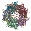 2iu7C 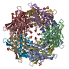 2iuoC  2iv1C  2ivgC 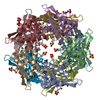 2ivqC  1dw9S S: Starting model for refinement C: citing same article ( |
|---|---|
| Similar structure data |


-Links
 PDBj
PDBj


- Assembly
| Deposited unit | 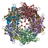
| ||||||||||||||||||||||||||||||||||||||||
|---|---|---|---|---|---|---|---|---|---|---|---|---|---|---|---|---|---|---|---|---|---|---|---|---|---|---|---|---|---|---|---|---|---|---|---|---|---|---|---|---|---|
| 1 |
| ||||||||||||||||||||||||||||||||||||||||
| Unit cell |
| ||||||||||||||||||||||||||||||||||||||||
| Noncrystallographic symmetry (NCS) | NCS oper:
| ||||||||||||||||||||||||||||||||||||||||
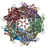
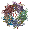
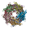
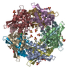
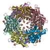
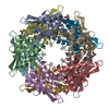
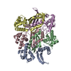
| Details | THE ENZYME IS A DECAMER MADE UP OF 5 DIMERS. 4 SUBUNITSCONTRIBUTE TO MAKING UP THE 5 ACTIVE SITES OF THISDECAMERIC ENZYME.THE DECAMER COULD BE VISUALIZED AS JI/CH/GE/FB/DA DIMERS. |
-Components
| #1: Protein | Mass: 17051.793 Da / Num. of mol.: 10 / Mutation: YES Source method: isolated from a genetically manipulated source Source: (gene. exp.) #2: Chemical | ChemComp-SO4 /   Mass: 96.063 Da / Num. of mol.: 16 / Source method: obtained synthetically / Formula: SO4 Mass: 96.063 Da / Num. of mol.: 16 / Source method: obtained synthetically / Formula: SO4#3: Chemical | ChemComp-CL /   Mass: 35.453 Da / Num. of mol.: 10 / Source method: obtained synthetically / Formula: Cl Mass: 35.453 Da / Num. of mol.: 10 / Source method: obtained synthetically / Formula: Cl#4: Chemical | ChemComp-AZI /   Mass: 42.020 Da / Num. of mol.: 5 / Source method: obtained synthetically / Formula: N3 Mass: 42.020 Da / Num. of mol.: 5 / Source method: obtained synthetically / Formula: N3#5: Water | ChemComp-HOH / |  Mass: 18.015 Da / Num. of mol.: 2728 / Source method: isolated from a natural source / Formula: H2O Mass: 18.015 Da / Num. of mol.: 2728 / Source method: isolated from a natural source / Formula: H2OCompound details | CATALYZES THE REACTION OF CYANATE WITH BICARBONATE TO PRODUCE AMMONIA AND CARBON DIOXIDE. ...CATALYZES THE REACTION OF CYANATE WITH BICARBONAT | Sequence details | SERINE 122 MUTATED TO ALANINE | |
|---|
-Experimental details
-Experiment
| Experiment | Method: X-RAY DIFFRACTION / Number of used crystals: 1 |
|---|
- Sample preparation
| Crystal | Density Matthews: 2.67 Å3/Da / Density % sol: 54 % / Description: NONE |
|---|---|
| Crystal grow | Method: vapor diffusion, sitting drop / pH: 7.3 Details: CRYSTALS WERE GROWN BY THE SITTING DROP METHOD OF VAPOUR DIFFUSION FROM 50% AMMONIUM SULPHATE SOLUTIONS BUFFERED WITH 50MM NAKPO4, PH 7.3, AND IN THE PRESENCE OF 50 MM TRIC/HCL, PH =7.3. |
-Data collection
| Diffraction | Mean temperature: 100 K |
|---|---|
| Diffraction source | Source: SYNCHROTRON / Site: APS  / Beamline: 19-ID / Wavelength: 1.033 / Beamline: 19-ID / Wavelength: 1.033 |
| Detector | Type: ADSC CCD / Detector: CCD / Details: MIRRORS |
| Radiation | Monochromator: SI 111 / Protocol: SINGLE WAVELENGTH / Monochromatic (M) / Laue (L): M / Scattering type: x-ray |
| Radiation wavelength | Wavelength: 1.033 Å / Relative weight: 1 |
| Reflection | Resolution: 1.95→75 Å / Num. obs: 115164 / % possible obs: 95.7 % / Redundancy: 2 % / Rmerge(I) obs: 0.04 / Net I/σ(I): 17.3 |
| Reflection shell | Resolution: 1.95→2.05 Å / Redundancy: 1.9 % / Rmerge(I) obs: 0.2 / Mean I/σ(I) obs: 5.3 / % possible all: 84.6 |
- Processing
| Software |
| ||||||||||||||||||||||||||||||||||||||||||||||||||||||||||||||||||||||||||||||||||||||||||||||||||||||||||||||||||||||||||||||||||||||||||||||||||||||||||||||||||||||||||||||||||||||
|---|---|---|---|---|---|---|---|---|---|---|---|---|---|---|---|---|---|---|---|---|---|---|---|---|---|---|---|---|---|---|---|---|---|---|---|---|---|---|---|---|---|---|---|---|---|---|---|---|---|---|---|---|---|---|---|---|---|---|---|---|---|---|---|---|---|---|---|---|---|---|---|---|---|---|---|---|---|---|---|---|---|---|---|---|---|---|---|---|---|---|---|---|---|---|---|---|---|---|---|---|---|---|---|---|---|---|---|---|---|---|---|---|---|---|---|---|---|---|---|---|---|---|---|---|---|---|---|---|---|---|---|---|---|---|---|---|---|---|---|---|---|---|---|---|---|---|---|---|---|---|---|---|---|---|---|---|---|---|---|---|---|---|---|---|---|---|---|---|---|---|---|---|---|---|---|---|---|---|---|---|---|---|---|
| Refinement | Method to determine structure: MOLECULAR REPLACEMENT Starting model: PDB ENTRY 1DW9 Resolution: 1.95→75.38 Å / Cor.coef. Fo:Fc: 0.967 / Cor.coef. Fo:Fc free: 0.933 / SU B: 3.365 / SU ML: 0.1 / Cross valid method: THROUGHOUT / ESU R: 0.16 / ESU R Free: 0.154 / Stereochemistry target values: MAXIMUM LIKELIHOOD / Details: HYDROGENS HAVE BEEN ADDED IN THE RIDING POSITIONS.
| ||||||||||||||||||||||||||||||||||||||||||||||||||||||||||||||||||||||||||||||||||||||||||||||||||||||||||||||||||||||||||||||||||||||||||||||||||||||||||||||||||||||||||||||||||||||
| Solvent computation | Ion probe radii: 0.8 Å / Shrinkage radii: 0.8 Å / VDW probe radii: 1.4 Å / Solvent model: MASK | ||||||||||||||||||||||||||||||||||||||||||||||||||||||||||||||||||||||||||||||||||||||||||||||||||||||||||||||||||||||||||||||||||||||||||||||||||||||||||||||||||||||||||||||||||||||
| Displacement parameters | Biso mean: 17.63 Å2
| ||||||||||||||||||||||||||||||||||||||||||||||||||||||||||||||||||||||||||||||||||||||||||||||||||||||||||||||||||||||||||||||||||||||||||||||||||||||||||||||||||||||||||||||||||||||
| Refinement step | Cycle: LAST / Resolution: 1.95→75.38 Å
| ||||||||||||||||||||||||||||||||||||||||||||||||||||||||||||||||||||||||||||||||||||||||||||||||||||||||||||||||||||||||||||||||||||||||||||||||||||||||||||||||||||||||||||||||||||||
| Refine LS restraints |
|