 Movie
Movie Controller

[English] 日本語
 Yorodumi
- PDB-2iuo: Site Directed Mutagenesis of Key Residues Involved in the Catalyt... -
+ Open data

- Basic information
| Entry | Database: PDB / ID: 2iuo | ||||||
|---|---|---|---|---|---|---|---|
| Title | Site Directed Mutagenesis of Key Residues Involved in the Catalytic Mechanism of Cyanase | ||||||
 Components Components | CYANATE HYDRATASE | ||||||
 Keywords Keywords | LYASE / CYANATE DEGRADATION | ||||||
| Function / homology |  Function and homology information Function and homology informationcyanase / cyanate hydratase activity / cyanate catabolic process / DNA binding Similarity search - Function | ||||||
| Biological species |  | ||||||
| Method |  X-RAY DIFFRACTION / SYNCHROTRON / MOLECULAR REPLACEMENT / Resolution: 1.9 Å X-RAY DIFFRACTION / SYNCHROTRON / MOLECULAR REPLACEMENT / Resolution: 1.9 Å | ||||||
 Authors Authors | Guilloton, M. / Walsh, M.A. / Joachimiak, A. / Anderson, M.P. | ||||||
 Citation Citation | Journal: To be Published Title: A Twin Set of Low Pka Arginines Ensures the Concerted Acid Base Catalytic Mechanism of Cyanase Authors: Guilloton, M. / Walsh, M.A. / Joachimiak, A. / Anderson, P.M. #1: Journal: Structure / Year: 2000Title: Structure of Cyanase Reveals that a Novel Dimeric and Decameric Arrangement of Subunits is Required for Formation of the Enzyme Active Site. Authors: Walsh, M.A. / Otwinowski, Z. / Perrakis, A. / Anderson, P.M. / Joachimiak, A. | ||||||
| History |
|
- Structure visualization
| Structure viewer | Molecule: MolmilJmol/JSmol |
|---|
- Downloads & links
-Download
| PDBx/mmCIF format | 2iuo.cif.gz | 348.7 KB | Display | PDBx/mmCIF format |
|---|---|---|---|---|
| PDB format | pdb2iuo.ent.gz | 285.2 KB | Display | PDB format |
| PDBx/mmJSON format | 2iuo.json.gz | Tree view | PDBx/mmJSON format | |
| Others |  Other downloads Other downloads |
-Validation report
| Arichive directory | https://data.pdbj.org/pub/pdb/validation_reports/iu/2iuoftp://data.pdbj.org/pub/pdb/validation_reports/iu/2iuo | HTTPS FTP |
|---|
-Related structure data
| Related structure data | 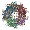 2iu7C  2iv1C 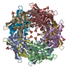 2ivbC  2ivgC 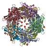 2ivqC  1dw9S S: Starting model for refinement C: citing same article ( |
|---|---|
| Similar structure data |


-Links
 PDBj
PDBj


- Assembly
| Deposited unit | 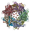
| ||||||||||||||||||||||||||||||||||||||||
|---|---|---|---|---|---|---|---|---|---|---|---|---|---|---|---|---|---|---|---|---|---|---|---|---|---|---|---|---|---|---|---|---|---|---|---|---|---|---|---|---|---|
| 1 |
| ||||||||||||||||||||||||||||||||||||||||
| Unit cell |
| ||||||||||||||||||||||||||||||||||||||||
| Noncrystallographic symmetry (NCS) | NCS oper:
| ||||||||||||||||||||||||||||||||||||||||
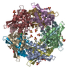
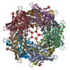
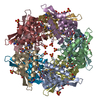
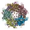
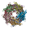
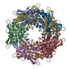
| Details | THE ENZYME IS A DECAMER MADE UO OF 5 DIMERS. 4 SUBUNITSCONTRIBUTE TO MAKING UP THE 5 ACTIVE SITES OF THISDECAMERIC ENZYME.THE DECAMER COULD BE VISUALIZED AS JI/CH/GE/FB/DA DIMERS. |
-Components
-Protein , 1 types, 10 molecules ABCDEFGHIJ
| #1: Protein | Mass: 17037.768 Da / Num. of mol.: 10 / Mutation: YES Source method: isolated from a genetically manipulated source Source: (gene. exp.) |
|---|
-Non-polymers , 5 types, 2096 molecules 


| #2: Chemical | ChemComp-BR /  Mass: 79.904 Da / Num. of mol.: 10 / Source method: obtained synthetically / Formula: Br Mass: 79.904 Da / Num. of mol.: 10 / Source method: obtained synthetically / Formula: Br#3: Chemical | ChemComp-CL /  Mass: 35.453 Da / Num. of mol.: 10 / Source method: obtained synthetically / Formula: Cl Mass: 35.453 Da / Num. of mol.: 10 / Source method: obtained synthetically / Formula: Cl#4: Chemical | ChemComp-SO4 /  Mass: 96.063 Da / Num. of mol.: 23 / Source method: obtained synthetically / Formula: SO4 Mass: 96.063 Da / Num. of mol.: 23 / Source method: obtained synthetically / Formula: SO4#5: Chemical | ChemComp-AZI / |  Mass: 42.020 Da / Num. of mol.: 1 / Source method: obtained synthetically / Formula: N3 Mass: 42.020 Da / Num. of mol.: 1 / Source method: obtained synthetically / Formula: N3#6: Water | ChemComp-HOH / | Mass: 18.015 Da / Num. of mol.: 2052 / Source method: isolated from a natural source / Formula: H2O |
|---|
-Details
| Sequence details | SERINE 122 MUTATED TO GLYCINE |
|---|
-Experimental details
-Experiment
| Experiment | Method: X-RAY DIFFRACTION / Number of used crystals: 1 |
|---|
- Sample preparation
| Crystal | Density Matthews: 2.67 Å3/Da / Density % sol: 54 % |
|---|---|
| Crystal grow | Method: vapor diffusion, sitting drop / pH: 7.3 Details: CRYSTALS WERE GROWN BY THE SITTING DROP METHOD OF VAPOUR DIFFUSION FROM 50% AMMONIUM SULPHATE SOLUTIONS BUFFERED WITH 50MM NAKPO4, PH = 7.3, AND IN THE PRESENCE OF 50 MM TRIC/HCL, PH =7.3. |
-Data collection
| Diffraction | Mean temperature: 100 K |
|---|---|
| Diffraction source | Source: SYNCHROTRON / Site: APS  / Beamline: 19-ID / Wavelength: 1.033 / Beamline: 19-ID / Wavelength: 1.033 |
| Detector | Type: ADSC CCD / Detector: CCD / Details: MIRRORS |
| Radiation | Monochromator: SI 111 / Protocol: SINGLE WAVELENGTH / Monochromatic (M) / Laue (L): M / Scattering type: x-ray |
| Radiation wavelength | Wavelength: 1.033 Å / Relative weight: 1 |
| Reflection | Resolution: 1.85→40 Å / Num. obs: 129043 / % possible obs: 92.5 % / Redundancy: 2.2 % / Rmerge(I) obs: 0.05 / Net I/σ(I): 14 |
| Reflection shell | Resolution: 1.85→1.92 Å / Redundancy: 2.1 % / Rmerge(I) obs: 0.24 / Mean I/σ(I) obs: 2.5 / % possible all: 59.9 |
- Processing
| Software |
| ||||||||||||||||||||||||||||||||||||||||||||||||||||||||||||||||||||||||||||||||||||||||||||||||||||||||||||||||||||||||||||||||||||||||||||||||||||||||||||||||||||||||||||||||||||||
|---|---|---|---|---|---|---|---|---|---|---|---|---|---|---|---|---|---|---|---|---|---|---|---|---|---|---|---|---|---|---|---|---|---|---|---|---|---|---|---|---|---|---|---|---|---|---|---|---|---|---|---|---|---|---|---|---|---|---|---|---|---|---|---|---|---|---|---|---|---|---|---|---|---|---|---|---|---|---|---|---|---|---|---|---|---|---|---|---|---|---|---|---|---|---|---|---|---|---|---|---|---|---|---|---|---|---|---|---|---|---|---|---|---|---|---|---|---|---|---|---|---|---|---|---|---|---|---|---|---|---|---|---|---|---|---|---|---|---|---|---|---|---|---|---|---|---|---|---|---|---|---|---|---|---|---|---|---|---|---|---|---|---|---|---|---|---|---|---|---|---|---|---|---|---|---|---|---|---|---|---|---|---|---|
| Refinement | Method to determine structure: MOLECULAR REPLACEMENT Starting model: PDB ENTRY 1DW9 Resolution: 1.9→76.7 Å / Cor.coef. Fo:Fc: 0.964 / Cor.coef. Fo:Fc free: 0.933 / SU B: 3.098 / SU ML: 0.093 / Cross valid method: THROUGHOUT / ESU R: 0.141 / ESU R Free: 0.142 / Stereochemistry target values: MAXIMUM LIKELIHOOD / Details: HYDROGENS HAVE BEEN ADDED IN THE RIDING POSITIONS.
| ||||||||||||||||||||||||||||||||||||||||||||||||||||||||||||||||||||||||||||||||||||||||||||||||||||||||||||||||||||||||||||||||||||||||||||||||||||||||||||||||||||||||||||||||||||||
| Solvent computation | Ion probe radii: 0.8 Å / Shrinkage radii: 0.8 Å / VDW probe radii: 1.4 Å / Solvent model: MASK | ||||||||||||||||||||||||||||||||||||||||||||||||||||||||||||||||||||||||||||||||||||||||||||||||||||||||||||||||||||||||||||||||||||||||||||||||||||||||||||||||||||||||||||||||||||||
| Displacement parameters | Biso mean: 13.54 Å2
| ||||||||||||||||||||||||||||||||||||||||||||||||||||||||||||||||||||||||||||||||||||||||||||||||||||||||||||||||||||||||||||||||||||||||||||||||||||||||||||||||||||||||||||||||||||||
| Refinement step | Cycle: LAST / Resolution: 1.9→76.7 Å
| ||||||||||||||||||||||||||||||||||||||||||||||||||||||||||||||||||||||||||||||||||||||||||||||||||||||||||||||||||||||||||||||||||||||||||||||||||||||||||||||||||||||||||||||||||||||
| Refine LS restraints |
|