 Movie
Movie Controller

[English] 日本語
 Yorodumi
- PDB-2iny: Nanoporous Crystals of Chicken Embryo Lethal Orphan (CELO) Adenov... -
+ Open data

- Basic information
| Entry | Database: PDB / ID: 2iny | ||||||
|---|---|---|---|---|---|---|---|
| Title | Nanoporous Crystals of Chicken Embryo Lethal Orphan (CELO) Adenovirus Major Coat Protein, Hexon | ||||||
 Components Components | Hexon protein | ||||||
 Keywords Keywords | VIRAL PROTEIN / AVIAN ADENOVIRUS / CELO / MAJOR COAT PROTEIN / HEXON / CRYSTAL PACKING / NANOTECHNOLOGY / VIRAL JELLY ROLL | ||||||
| Function / homology |  Function and homology information Function and homology informationT=25 icosahedral viral capsid / microtubule-dependent intracellular transport of viral material towards nucleus / host cell / symbiont entry into host cell / host cell nucleus / structural molecule activity Similarity search - Function | ||||||
| Biological species |  Fowl adenovirus 1 Fowl adenovirus 1 | ||||||
| Method |  X-RAY DIFFRACTION / MOLECULAR REPLACEMENT / Resolution: 3.9 Å X-RAY DIFFRACTION / MOLECULAR REPLACEMENT / Resolution: 3.9 Å | ||||||
 Authors Authors | Xu, L. / Benson, S.D. / Burnett, R.M. | ||||||
 Citation Citation | Journal: J.Struct.Biol. / Year: 2007 Title: Nanoporous crystals of chicken embryo lethal orphan (CELO) adenovirus major coat protein, hexon. Authors: Xu, L. / Benson, S.D. / Burnett, R.M. #1: Journal: J.Virol. / Year: 2003Title: Structural and Phylogenetic Analysis of Adenovirus Hexons by Use of High-Resolution X-Ray Crystallographic, Molecular Modeling, and Sequence-Based Methods Authors: Rux, J.J. / Kuser, P.R. / Burnett, R.M. #2: Journal: Mol.Ther. / Year: 2000Title: Type-Specific Epitope Locations Revealed by X-Ray Crystallographic Study of Adenovirus Type 5 Hexon Authors: Rux, J.J. / Burnett, R.M. #3: Journal: J.Struct.Biol. / Year: 2006Title: Capsid-Like Arrays in Crystals of Chimpanzee Adenovirus Hexon Authors: Xue, F. / Burnett, R.M. #4: Journal: Virology / Year: 1971Title: Purification and Properties of Chick Embryo Lethal Orphan Virus (an Avian Adenovirus) Authors: Laver, W.G. / Younghusband, H.B. / Wrigley, N.G. #5: Journal: J.Virol. / Year: 1996Title: The Complete DNA Sequence and Genomic Organization of the Avian Adenovirus CELO Authors: Chiocca, S. / Kurzbauer, R. / Schaffner, G. / Baker, A. / Mautner, V. / Cotten, M. #6: Journal: J.Virol. / Year: 1999Title: Mutational Analysis of the Avian Adenovirus CELO, which Provides a Basis for Gene Delivery Vectors Authors: Michou, A.I. / Lehrmann, H. / Saltik, M. / Cotten, M. #7: Journal: J.Virol. / Year: 1993Title: Chicken Adenovirus (CELO Virus) Particles Augment Receptor-Mediated DNA Delivery to Mammalian Cells and Yield Exceptional Levels of Stable Transformants Authors: Cotten, M. / Wagner, E. / Zatloukal, K. / Birnstiel, M.L. #8: Journal: J.Virol. / Year: 1996Title: Analysis of 15 Adenovirus Hexon Proteins Reveals the Location and Structure of Seven Hypervariable Regions Containing Serotype-Specific Residues Authors: Crawford-Miksza, L. / Schnurr, D.P. | ||||||
| History |
|
- Structure visualization
| Structure viewer | Molecule: MolmilJmol/JSmol |
|---|
- Downloads & links
-Download
| PDBx/mmCIF format | 2iny.cif.gz | 180.3 KB | Display | PDBx/mmCIF format |
|---|---|---|---|---|
| PDB format | pdb2iny.ent.gz | 135.9 KB | Display | PDB format |
| PDBx/mmJSON format | 2iny.json.gz | Tree view | PDBx/mmJSON format | |
| Others |  Other downloads Other downloads |
-Validation report
| Arichive directory | https://data.pdbj.org/pub/pdb/validation_reports/in/2inyftp://data.pdbj.org/pub/pdb/validation_reports/in/2iny | HTTPS FTP |
|---|
-Related structure data
| Related structure data |  1p30S S: Starting model for refinement |
|---|---|
| Similar structure data |


-Links
 PDBj
PDBj


- Assembly
| Deposited unit | 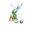
| ||||||||
|---|---|---|---|---|---|---|---|---|---|
| 1 | 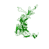
| ||||||||
| Unit cell |
| ||||||||
| Details | The biological assembly is a trimer generated from the monmer in the asymmetric unit by the operators: -y, x-y+1, z and -x+y-1, -x, z |
-Components
| #1: Protein | Mass: 106795.633 Da / Num. of mol.: 1 / Source method: isolated from a natural source / Source: (natural) Fowl adenovirus 1 / Genus: Aviadenovirus / Species: Fowl adenovirus A / Strain: Phelps Strain / References: UniProt: P42671 |
|---|
-Experimental details
-Experiment
| Experiment | Method: X-RAY DIFFRACTION / Number of used crystals: 1 |
|---|
- Sample preparation
| Crystal | Density Matthews: 3.84 Å3/Da / Density % sol: 67.97 % |
|---|---|
| Crystal grow | Temperature: 295 K / Method: vapor diffusion, hanging drop / pH: 7.5 Details: 7% iso-propanol, 13% PEG 4000, 0.01 M glycine, 0.1 M Hepes, pH 7.5, VAPOR DIFFUSION, HANGING DROP, temperature 295.0K |
-Data collection
| Diffraction | Mean temperature: 298 K |
|---|---|
| Diffraction source | Source: ROTATING ANODE / Type: RIGAKU RU200 / Wavelength: 1.5418 |
| Detector | Type: RIGAKU RAXIS II / Detector: IMAGE PLATE / Details: mirrors |
| Radiation | Monochromator: YALE MIRRORS / Protocol: SINGLE WAVELENGTH / Monochromatic (M) / Laue (L): M / Scattering type: x-ray |
| Radiation wavelength | Wavelength: 1.5418 Å / Relative weight: 1 |
| Reflection | Resolution: 3.9→50 Å / Num. all: 15294 / Num. obs: 14636 / % possible obs: 95.7 % / Observed criterion σ(I): 1 / Redundancy: 4.6 % / Rsym value: 0.205 / Net I/σ(I): 7.1 |
| Reflection shell | Resolution: 3.9→3.97 Å / Redundancy: 4.2 % / Mean I/σ(I) obs: 2.5 / Num. unique all: 712 / Rsym value: 0.478 / % possible all: 94.4 |
- Processing
| Software |
| ||||||||||||||||||||||||||||
|---|---|---|---|---|---|---|---|---|---|---|---|---|---|---|---|---|---|---|---|---|---|---|---|---|---|---|---|---|---|
| Refinement | Method to determine structure: MOLECULAR REPLACEMENT Starting model: PDB Entry 1P30 Resolution: 3.9→47.06 Å / Rfactor Rfree error: 0.016 / Data cutoff high absF: 154406.875 / Data cutoff low absF: 0 / Isotropic thermal model: OVERALL / Cross valid method: THROUGHOUT / σ(F): 0 / Stereochemistry target values: Engh & Huber Details: The structure was determined by molecular replacement using a model based on a threading of the CELO hexon sequence onto the human adenovirus type 5 hexon structure (1P30) using the program ...Details: The structure was determined by molecular replacement using a model based on a threading of the CELO hexon sequence onto the human adenovirus type 5 hexon structure (1P30) using the program JACKAL; The residues that are consistent with the electron density are defined with an occupancy of 1, the areas that are disordered are designated with an occupancy of 0.
| ||||||||||||||||||||||||||||
| Solvent computation | Solvent model: FLAT MODEL / Bsol: 110.542 Å2 / ksol: 0.076 e/Å3 | ||||||||||||||||||||||||||||
| Displacement parameters | Biso mean: 2.8 Å2
| ||||||||||||||||||||||||||||
| Refine analyze |
| ||||||||||||||||||||||||||||
| Refinement step | Cycle: LAST / Resolution: 3.9→47.06 Å
| ||||||||||||||||||||||||||||
| Refine LS restraints |
| ||||||||||||||||||||||||||||
| LS refinement shell | Resolution: 3.9→4.14 Å / Rfactor Rfree error: 0.045 / Total num. of bins used: 6
| ||||||||||||||||||||||||||||
| Xplor file |
|
