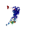
 Movie
Movie Controller


+ Open data

- Basic information
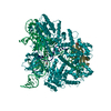
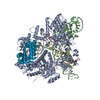
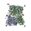
| Entry | Database: PDB / ID: 1p2z | |||||||||
|---|---|---|---|---|---|---|---|---|---|---|
| Title | Refinement of Adenovirus Type 2 Hexon with CNS | |||||||||
 Components Components | Hexon protein | |||||||||
 Keywords Keywords | VIRAL PROTEIN / ADENOVIRUS / TYPE 2 / HEXON / VIRUS / JELLYROLL / COAT PROTEIN | |||||||||
| Function / homology |  Function and homology information Function and homology informationT=25 icosahedral viral capsid / microtubule-dependent intracellular transport of viral material towards nucleus / host cell / symbiont entry into host cell / host cell nucleus / structural molecule activity Similarity search - Function | |||||||||
| Biological species |   Human adenovirus 2 Human adenovirus 2 | |||||||||
| Method |  X-RAY DIFFRACTION / MOLECULAR REPLACEMENT / Resolution: 2.2 Å X-RAY DIFFRACTION / MOLECULAR REPLACEMENT / Resolution: 2.2 Å | |||||||||
 Authors Authors | Rux, J.J. / Kuser, P.R. / Burnett, R.M. | |||||||||
 Citation Citation | Journal: J.VIROL. / Year: 2003 Title: Structural and phylogenetic analysis of adenovirus hexons by use of high-resolution x-ray crystallographic, molecular modeling, and sequence-based methods Authors: Rux, J.J. / Kuser, P.R. / Burnett, R.M. #1: Journal: MOL.THER. / Year: 2000Title: Type-specific epitope locations revealed by X-ray crystallographic study of adenovirus type 5 hexon. Authors: Rux, J.J. / Burnett, R.M. #2: Journal: J.Mol.Biol. / Year: 1994Title: The refined crystal structure of hexon, the major coat protein of adenovirus type 2, at 2.9 A resolution. Authors: Athappilly, F.K. / Murali, R. / Rux, J.J. / Cai, Z. / Burnett, R.M. #3: Journal: ADENOVIRUS METHODS AND PROTOCOLS (IN: METHODS IN MOLECULAR MEDICINE, V.21)Year: 1999 Title: Large-scale purification and crystallization of adenovirus hexon Authors: Rux, J.J. / Pascolini, D. / Burnett, R.M. | |||||||||
| History |
| |||||||||
| Remark 999 | SEQUENCE THE PROTEIN IS ACETYLATED AT THE N-TERMINUS. |
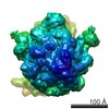
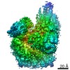
- Structure visualization
| Structure viewer | Molecule: MolmilJmol/JSmol |
|---|
- Downloads & links
-Download
| PDBx/mmCIF format | 1p2z.cif.gz | 196.4 KB | Display | PDBx/mmCIF format |
|---|---|---|---|---|
| PDB format | pdb1p2z.ent.gz | 153.6 KB | Display | PDB format |
| PDBx/mmJSON format | 1p2z.json.gz | Tree view | PDBx/mmJSON format | |
| Others |  Other downloads Other downloads |
-Validation report
| Arichive directory | https://data.pdbj.org/pub/pdb/validation_reports/p2/1p2zftp://data.pdbj.org/pub/pdb/validation_reports/p2/1p2z | HTTPS FTP |
|---|
-Related structure data
| Related structure data |  1p30C  1rux C: citing same article ( S: Starting model for refinement |
|---|---|
| Similar structure data |

-Links
 PDBj
PDBj


- Assembly
| Deposited unit | 
| |||||||||
|---|---|---|---|---|---|---|---|---|---|---|
| 1 | 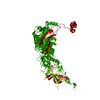
| |||||||||
| Unit cell |
| |||||||||
| Components on special symmetry positions |
| |||||||||
| Details | The biological assembly is a trimer generated by the operations z, x, y and y, z, x. |
-Components
| #1: Protein | Mass: 109121.133 Da / Num. of mol.: 1 / Source method: isolated from a natural source / Source: (natural) Human adenovirus 2 / Genus: Mastadenovirus / Species: Human adenovirus C / References: UniProt: P03277 | ||
|---|---|---|---|
| #2: Chemical | ChemComp-CIT /   Mass: 192.124 Da / Num. of mol.: 5 / Source method: obtained synthetically / Formula: C6H8O7 Mass: 192.124 Da / Num. of mol.: 5 / Source method: obtained synthetically / Formula: C6H8O7#3: Water | ChemComp-HOH / |  Mass: 18.015 Da / Num. of mol.: 430 / Source method: isolated from a natural source / Formula: H2O Mass: 18.015 Da / Num. of mol.: 430 / Source method: isolated from a natural source / Formula: H2O |
-Experimental details
-Experiment
| Experiment | Method: X-RAY DIFFRACTION / Number of used crystals: 1 |
|---|
- Sample preparation
| Crystal | Density Matthews: 2.61 Å3/Da / Density % sol: 52.8 % |
|---|---|
| Crystal grow | Temperature: 298 K / Method: vapor diffusion, hanging drop / pH: 3.2 Details: 0.5 M SODIUM CITRATE , pH 3.2, VAPOR DIFFUSION, HANGING DROP, temperature 298K |
-Data collection
| Diffraction | Mean temperature: 298 K |
|---|---|
| Diffraction source | Source: ROTATING ANODE / Type: RIGAKU RU200 / Wavelength: 1.5418 Å |
| Detector | Type: SIEMENS-NICOLET X100 / Detector: AREA DETECTOR / Date: Apr 1, 1991 / Details: MIRRORS |
| Radiation | Monochromator: double-mirror focusing system (Supper) / Protocol: SINGLE WAVELENGTH / Monochromatic (M) / Laue (L): M / Scattering type: x-ray |
| Radiation wavelength | Wavelength: 1.5418 Å / Relative weight: 1 |
| Reflection | Resolution: 2.2→10 Å / Num. all: 57118 / Num. obs: 48199 / % possible obs: 84.4 % / Observed criterion σ(F): 0 / Observed criterion σ(I): 0 / Biso Wilson estimate: 5.6 Å2 / Rsym value: 0.098 |
| Reflection shell | Resolution: 2.2→2.32 Å / Num. unique all: 7162 / % possible all: 75.8 |
- Processing
| Software |
| ||||||||||||||||||||||||||||||||||||
|---|---|---|---|---|---|---|---|---|---|---|---|---|---|---|---|---|---|---|---|---|---|---|---|---|---|---|---|---|---|---|---|---|---|---|---|---|---|
| Refinement | Method to determine structure: MOLECULAR REPLACEMENT Starting model: PDB ENTRY 1RUX 1rux Resolution: 2.2→9.99 Å / Rfactor Rfree error: 0.004 / Isotropic thermal model: RESTRAINED / Cross valid method: THROUGHOUT / σ(F): 0 / Stereochemistry target values: Engh & Huber
| ||||||||||||||||||||||||||||||||||||
| Solvent computation | Solvent model: FLAT MODEL / Bsol: 67.9291 Å2 / ksol: 0.399606 e/Å3 | ||||||||||||||||||||||||||||||||||||
| Displacement parameters | Biso mean: 18.8 Å2
| ||||||||||||||||||||||||||||||||||||
| Refine analyze |
| ||||||||||||||||||||||||||||||||||||
| Refinement step | Cycle: LAST / Resolution: 2.2→9.99 Å
| ||||||||||||||||||||||||||||||||||||
| Refine LS restraints |
| ||||||||||||||||||||||||||||||||||||
| LS refinement shell | Resolution: 2.2→2.34 Å / Rfactor Rfree error: 0.016 / Total num. of bins used: 6
| ||||||||||||||||||||||||||||||||||||
| Xplor file |
|
