Resolution: 2.4→2.5 Å / Redundancy: 3 % / Rmerge(I) obs: 0.427 / Mean I/σ(I) obs: 3.2 / Num. unique all: 19000 / % possible all: 99.2
-
Processing
Software
Name
Version
Classification
PHASER
phasing
REFMAC
5.5.0102
refinement
XDS
datareduction
XSCALE
datascaling
Refinement
Method to determine structure: FOURIER SYNTHESIS / Resolution: 2.4→20 Å / Cor.coef. Fo:Fc: 0.915 / Cor.coef. Fo:Fc free: 0.873 / SU B: 8.979 / SU ML: 0.209 / Cross valid method: THROUGHOUT / σ(F): 0 / σ(I): 0 / ESU R Free: 0.28 / Stereochemistry target values: MAXIMUM LIKELIHOOD Details: HYDROGENS HAVE BEEN ADDED IN THE RIDING POSITIONS. INITIAL MODEL OBTAINED WITH SELENIUM-SAD ON THE SAME CRYSTAL FORM
Rfactor
Num. reflection
% reflection
Selection details
Rfree
0.27091
9136
5.6 %
THIN RESOLUTION SHELLS DUE TO CRYSTALLOGRAPHIC SYMMETRY
Rwork
0.22386
-
-
-
all
0.22648
154688
-
-
obs
0.22648
154688
98.16 %
-
Solvent computation
Ion probe radii: 0.8 Å / Shrinkage radii: 0.8 Å / VDW probe radii: 1.4 Å / Solvent model: BABINET MODEL WITH MASK
Displacement parameters
Biso mean: 31.246 Å2
Baniso -1
Baniso -2
Baniso -3
1-
0.09 Å2
0 Å2
-0.17 Å2
2-
-
1.04 Å2
0 Å2
3-
-
-
-1.13 Å2
Refinement step
Cycle: LAST / Resolution: 2.4→20 Å
Protein
Nucleic acid
Ligand
Solvent
Total
Num. atoms
26297
0
84
840
27221
Refine LS restraints
Refine-ID
Type
Dev ideal
Dev ideal target
Number
X-RAY DIFFRACTION
r_bond_refined_d
0.005
0.022
26917
X-RAY DIFFRACTION
r_bond_other_d
0.001
0.02
18863
X-RAY DIFFRACTION
r_angle_refined_deg
0.841
1.966
36368
X-RAY DIFFRACTION
r_angle_other_deg
0.767
3.001
45706
X-RAY DIFFRACTION
r_dihedral_angle_1_deg
4.977
5
3215
X-RAY DIFFRACTION
r_dihedral_angle_2_deg
32.478
23.457
1296
X-RAY DIFFRACTION
r_dihedral_angle_3_deg
12.652
15
4895
X-RAY DIFFRACTION
r_dihedral_angle_4_deg
11.027
15
224
X-RAY DIFFRACTION
r_chiral_restr
0.05
0.2
4046
X-RAY DIFFRACTION
r_gen_planes_refined
0.003
0.021
29385
X-RAY DIFFRACTION
r_gen_planes_other
0.001
0.02
5551
X-RAY DIFFRACTION
r_mcbond_it
0.232
1.5
16091
X-RAY DIFFRACTION
r_mcbond_other
0.025
1.5
6494
X-RAY DIFFRACTION
r_mcangle_it
0.448
2
26279
X-RAY DIFFRACTION
r_scbond_it
0.581
3
10826
X-RAY DIFFRACTION
r_scangle_it
0.973
4.5
10089
LS refinement shell
Resolution: 2.4→2.462 Å / Total num. of bins used: 20
Rfactor
Num. reflection
% reflection
Rwork
0.289
12249
-
Rfree
-
0
-
obs
-
-
99.16 %
+
About Yorodumi
-
News
-
Feb 9, 2022. New format data for meta-information of EMDB entries
New format data for meta-information of EMDB entries
Version 3 of the EMDB header file is now the official format.
The previous official version 1.9 will be removed from the archive.
In the structure databanks used in Yorodumi, some data are registered as the other names, "COVID-19 virus" and "2019-nCoV". Here are the details of the virus and the list of structure data.
Jan 31, 2019. EMDB accession codes are about to change! (news from PDBe EMDB page)
EMDB accession codes are about to change! (news from PDBe EMDB page)
The allocation of 4 digits for EMDB accession codes will soon come to an end. Whilst these codes will remain in use, new EMDB accession codes will include an additional digit and will expand incrementally as the available range of codes is exhausted. The current 4-digit format prefixed with “EMD-” (i.e. EMD-XXXX) will advance to a 5-digit format (i.e. EMD-XXXXX), and so on. It is currently estimated that the 4-digit codes will be depleted around Spring 2019, at which point the 5-digit format will come into force.
The EM Navigator/Yorodumi systems omit the EMD- prefix.
Related info.:Q: What is EMD? / ID/Accession-code notation in Yorodumi/EM Navigator
Yorodumi is a browser for structure data from EMDB, PDB, SASBDB, etc.
This page is also the successor to EM Navigator detail page, and also detail information page/front-end page for Omokage search.
The word "yorodu" (or yorozu) is an old Japanese word meaning "ten thousand". "mi" (miru) is to see.
Related info.:EMDB / PDB / SASBDB / Comparison of 3 databanks / Yorodumi Search / Aug 31, 2016. New EM Navigator & Yorodumi / Yorodumi Papers / Jmol/JSmol / Function and homology information / Changes in new EM Navigator and Yorodumi
 Movie
Movie Controller


 Open data
 Basic information
 Components
 Keywords
 Function and homology information
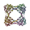
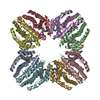
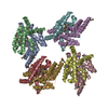
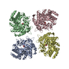
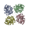
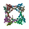
 Acidianus sp. A1-3 (archaea)
 X-RAY DIFFRACTION /
  Authors
 Citation
 Structure visualization
 Downloads & links
 Other downloads
 PDBj
PDBj
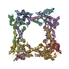
 Assembly
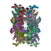


 Mass: 35.453 Da / Num. of mol.: 22 / Source method: obtained synthetically / Formula: Cl
 Mass: 634.751 Da / Num. of mol.: 2 / Source method: obtained synthetically / Formula: C28H58O15
 Mass: 18.015 Da / Num. of mol.: 840 / Source method: isolated from a natural source / Formula: H2O
 Sample preparation
 / Beamline: X10SA / Wavelength: 0.98089 Å
 Processing