 Movie
Movie Controller

[English] 日本語
 Yorodumi
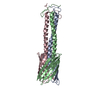
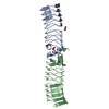
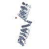
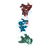
- PDB-2ibl: Crystal structure of a helper molecule (HT-mf-thromb) based on mi... -
+ Open data

- Basic information
| Entry | Database: PDB / ID: 2ibl | ||||||
|---|---|---|---|---|---|---|---|
| Title | Crystal structure of a helper molecule (HT-mf-thromb) based on mini-fibritin (mf) crystal structure (pdb:1OX3). | ||||||
 Components Components | Fibritin | ||||||
 Keywords Keywords | CHAPERONE / mini-fibritin / foldon / trimerization / bacteriophage t4 / helper molecule | ||||||
| Function / homology | 6-Phosphogluconate Dehydrogenase, domain 3 / Fibritin C-terminal / Fibritin C-terminal region / Single alpha-helices involved in coiled-coils or other helix-helix interfaces / virion component / Up-down Bundle / Mainly Alpha / Fibritin / Fibritin Function and homology information Function and homology information | ||||||
| Biological species |  Enterobacteria phage T4 (virus)Enterobacteria phage Ox2 (virus) Enterobacteria phage T4 (virus)Enterobacteria phage Ox2 (virus) | ||||||
| Method |  X-RAY DIFFRACTION / SYNCHROTRON / MOLECULAR REPLACEMENT / Resolution: 1.32 Å X-RAY DIFFRACTION / SYNCHROTRON / MOLECULAR REPLACEMENT / Resolution: 1.32 Å | ||||||
 Authors Authors | Boudko, S.P. / Rossmann, M.G. | ||||||
 Citation Citation | Journal: J.Mol.Biol. / Year: 2007 Title: The Coiled-coil Domain Structure of the Sin Nombre Virus Nucleocapsid Protein. Authors: Boudko, S.P. / Kuhn, R.J. / Rossmann, M.G. #1: Journal: J.Mol.Biol. / Year: 2004Title: Structure formation in the C terminus of type III collagen guides disulfide cross-linking Authors: Boudko, S.P. / Engel, J. #2: Journal: J.Mol.Biol. / Year: 2004Title: Design and crystal structure of bacteriophage T4 mini-fibritin NCCF Authors: Boudko, S.P. / Strelkov, S.V. / Engel, J. / Stetefeld, J. | ||||||
| History |
|
- Structure visualization
| Structure viewer | Molecule: MolmilJmol/JSmol |
|---|
- Downloads & links
-Download
| PDBx/mmCIF format | 2ibl.cif.gz | 59.7 KB | Display | PDBx/mmCIF format |
|---|---|---|---|---|
| PDB format | pdb2ibl.ent.gz | 42.8 KB | Display | PDB format |
| PDBx/mmJSON format | 2ibl.json.gz | Tree view | PDBx/mmJSON format | |
| Others |  Other downloads Other downloads |
-Validation report
| Arichive directory | https://data.pdbj.org/pub/pdb/validation_reports/ib/2iblftp://data.pdbj.org/pub/pdb/validation_reports/ib/2ibl | HTTPS FTP |
|---|
-Related structure data
| Related structure data |  2ic6C 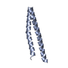 2ic9C  1ox3S S: Starting model for refinement C: citing same article ( |
|---|---|
| Similar structure data |


-Links
 PDBj
PDBj


- Assembly
| Deposited unit | 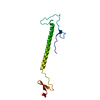
| ||||||||||||
|---|---|---|---|---|---|---|---|---|---|---|---|---|---|
| 1 | 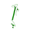
| ||||||||||||
| Unit cell |
| ||||||||||||
| Components on special symmetry positions |
| ||||||||||||
| Details | The biological assembly is a trimer generated from the monomer in the asymmetric unit by the operations: x,y,z; -x+y+1,-x,z; -y,x-y-1,z |
-Components
| #1: Protein | Mass: 14061.588 Da / Num. of mol.: 1 Source method: isolated from a genetically manipulated source Source: (gene. exp.) Enterobacteria phage T4 (virus), (gene. exp.) Enterobacteria phage Ox2 (virus)Gene: wac / Plasmid: pET23d(+) / Species (production host): Escherichia coli / Production host:  |
|---|---|
| #2: Water | ChemComp-HOH /  Mass: 18.015 Da / Num. of mol.: 197 / Source method: isolated from a natural source / Formula: H2O Mass: 18.015 Da / Num. of mol.: 197 / Source method: isolated from a natural source / Formula: H2O |
-Experimental details
-Experiment
| Experiment | Method: X-RAY DIFFRACTION / Number of used crystals: 1 |
|---|
- Sample preparation
| Crystal | Density Matthews: 3.15 Å3/Da / Density % sol: 60.91 % |
|---|---|
| Crystal grow | Temperature: 293 K / Method: vapor diffusion, hanging drop / pH: 10.5 Details: 30% PEG 400, 0.1 M TrisHCl, 0.2 M magnesium chloride, pH 10.5, VAPOR DIFFUSION, HANGING DROP, temperature 293.0K |
-Data collection
| Diffraction | Mean temperature: 100 K |
|---|---|
| Diffraction source | Source: SYNCHROTRON / Site: APS  / Beamline: 23-ID-D / Wavelength: 1.00465 Å / Beamline: 23-ID-D / Wavelength: 1.00465 Å |
| Detector | Type: MARMOSAIC 300 mm CCD / Detector: CCD / Date: Oct 30, 2005 Details: Si(111) Double Crystal Monochrometer. Adjustable focusing mirrors in K-B geometry |
| Radiation | Monochromator: double crystal monochromator and K-B pair of biomorph mirrors for vertical and horizontal focusing Protocol: SINGLE WAVELENGTH / Monochromatic (M) / Laue (L): M / Scattering type: x-ray |
| Radiation wavelength | Wavelength: 1.00465 Å / Relative weight: 1 |
| Reflection | Resolution: 1.32→35 Å / Num. all: 40437 / Num. obs: 40028 / % possible obs: 98.1 % / Observed criterion σ(F): 2 / Observed criterion σ(I): 2 / Redundancy: 6.6 % / Biso Wilson estimate: 15.7 Å2 / Rmerge(I) obs: 0.056 / Rsym value: 0.056 / Net I/σ(I): 31.7 |
| Reflection shell | Resolution: 1.32→1.354 Å / Redundancy: 4.2 % / Rmerge(I) obs: 0.216 / Mean I/σ(I) obs: 5.2 / Num. unique all: 2823 / Rsym value: 0.216 / % possible all: 89.71 |
- Processing
| Software |
| |||||||||||||||||||||||||||||||||||||||||||||||||||||||||||||||||||||||||||||||||||||||||||||||||||||||||||||||||||||||||||||
|---|---|---|---|---|---|---|---|---|---|---|---|---|---|---|---|---|---|---|---|---|---|---|---|---|---|---|---|---|---|---|---|---|---|---|---|---|---|---|---|---|---|---|---|---|---|---|---|---|---|---|---|---|---|---|---|---|---|---|---|---|---|---|---|---|---|---|---|---|---|---|---|---|---|---|---|---|---|---|---|---|---|---|---|---|---|---|---|---|---|---|---|---|---|---|---|---|---|---|---|---|---|---|---|---|---|---|---|---|---|---|---|---|---|---|---|---|---|---|---|---|---|---|---|---|---|---|
| Refinement | Method to determine structure: MOLECULAR REPLACEMENT Starting model: PDB ENTRY 1OX3 Resolution: 1.32→35 Å / Cor.coef. Fo:Fc: 0.97 / Cor.coef. Fo:Fc free: 0.969 / SU B: 0.972 / SU ML: 0.021 / Cross valid method: THROUGHOUT / σ(F): 0 / σ(I): 0 / ESU R: 0.041 / ESU R Free: 0.038 / Stereochemistry target values: MAXIMUM LIKELIHOOD / Details: HYDROGENS HAVE BEEN ADDED IN THE RIDING POSITIONS
| |||||||||||||||||||||||||||||||||||||||||||||||||||||||||||||||||||||||||||||||||||||||||||||||||||||||||||||||||||||||||||||
| Solvent computation | Ion probe radii: 0.8 Å / Shrinkage radii: 0.8 Å / VDW probe radii: 1.4 Å / Solvent model: BABINET MODEL WITH MASK | |||||||||||||||||||||||||||||||||||||||||||||||||||||||||||||||||||||||||||||||||||||||||||||||||||||||||||||||||||||||||||||
| Displacement parameters | Biso mean: 13.117 Å2
| |||||||||||||||||||||||||||||||||||||||||||||||||||||||||||||||||||||||||||||||||||||||||||||||||||||||||||||||||||||||||||||
| Refinement step | Cycle: LAST / Resolution: 1.32→35 Å
| |||||||||||||||||||||||||||||||||||||||||||||||||||||||||||||||||||||||||||||||||||||||||||||||||||||||||||||||||||||||||||||
| Refine LS restraints |
| |||||||||||||||||||||||||||||||||||||||||||||||||||||||||||||||||||||||||||||||||||||||||||||||||||||||||||||||||||||||||||||
| LS refinement shell | Resolution: 1.32→1.354 Å / Total num. of bins used: 20
|
