 Movie
Movie Controller

[English] 日本語
 Yorodumi
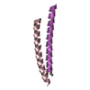
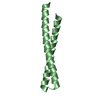
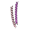
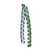
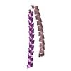
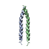
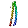
- PDB-2ic9: The Coiled-coil Domain (residues 1-93) Structure of the Sin Nombr... -
+ Open data

- Basic information
| Entry | Database: PDB / ID: 2ic9 | ||||||
|---|---|---|---|---|---|---|---|
| Title | The Coiled-coil Domain (residues 1-93) Structure of the Sin Nombre Virus Nucleocapsid Protein | ||||||
 Components Components | Nucleocapsid protein | ||||||
 Keywords Keywords | VIRAL PROTEIN / Sin Nombre Virus / Hantavirus / Bunyaviridae / ssRNA negative-strand viruses / nucleocapsid protein / antiparallel coiled coil | ||||||
| Function / homology |  Function and homology information Function and homology informationviral nucleocapsid / endonuclease activity / Hydrolases; Acting on ester bonds / host cell Golgi apparatus / host cell perinuclear region of cytoplasm / ribonucleoprotein complex / hydrolase activity / RNA binding Similarity search - Function | ||||||
| Biological species |  Sin Nombre virus Sin Nombre virus | ||||||
| Method |  X-RAY DIFFRACTION / SYNCHROTRON / MOLECULAR REPLACEMENT / Resolution: 2 Å X-RAY DIFFRACTION / SYNCHROTRON / MOLECULAR REPLACEMENT / Resolution: 2 Å | ||||||
 Authors Authors | Boudko, S.P. / Rossmann, M.G. | ||||||
 Citation Citation | Journal: J.Mol.Biol. / Year: 2007 Title: The Coiled-coil Domain Structure of the Sin Nombre Virus Nucleocapsid Protein. Authors: Boudko, S.P. / Kuhn, R.J. / Rossmann, M.G. | ||||||
| History |
|
- Structure visualization
| Structure viewer | Molecule: MolmilJmol/JSmol |
|---|
- Downloads & links
-Download
| PDBx/mmCIF format | 2ic9.cif.gz | 42.5 KB | Display | PDBx/mmCIF format |
|---|---|---|---|---|
| PDB format | pdb2ic9.ent.gz | 30.4 KB | Display | PDB format |
| PDBx/mmJSON format | 2ic9.json.gz | Tree view | PDBx/mmJSON format | |
| Others |  Other downloads Other downloads |
-Validation report
| Arichive directory | https://data.pdbj.org/pub/pdb/validation_reports/ic/2ic9ftp://data.pdbj.org/pub/pdb/validation_reports/ic/2ic9 | HTTPS FTP |
|---|
-Related structure data
| Related structure data | 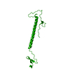 2iblC  2ic6SC S: Starting model for refinement C: citing same article ( |
|---|---|
| Similar structure data |


-Links
 PDBj
PDBj- Assembly
| Deposited unit | 
| ||||||||
|---|---|---|---|---|---|---|---|---|---|
| 1 | 
| ||||||||
| 2 | 
| ||||||||
| Unit cell |
|
-Components
| #1: Protein | Mass: 10700.089 Da / Num. of mol.: 2 / Fragment: Residues: 1-93 Source method: isolated from a genetically manipulated source Source: (gene. exp.) Sin Nombre virus / Genus: Hantavirus / Strain: Convict Creek 107 virus / Gene: nucleocapsid protein / Plasmid: pET23d(+) / Species (production host): Escherichia coli / Production host:  #2: Water | ChemComp-HOH / |  Mass: 18.015 Da / Num. of mol.: 92 / Source method: isolated from a natural source / Formula: H2O Mass: 18.015 Da / Num. of mol.: 92 / Source method: isolated from a natural source / Formula: H2O |
|---|
-Experimental details
-Experiment
| Experiment | Method: X-RAY DIFFRACTION / Number of used crystals: 1 |
|---|
- Sample preparation
| Crystal | Density Matthews: 2.12 Å3/Da / Density % sol: 42.01 % |
|---|---|
| Crystal grow | Temperature: 293 K / Method: vapor diffusion, hanging drop / pH: 7.5 Details: 0.15 M potassium bromide, 0.1 M Tris-HCl, 34% PEG mono-methyl ether (PEG MME) 200, pH 7.5, VAPOR DIFFUSION, HANGING DROP, temperature 293K |
-Data collection
| Diffraction | Mean temperature: 100 K |
|---|---|
| Diffraction source | Source: SYNCHROTRON / Site: APS  / Beamline: 14-BM-C / Wavelength: 0.9002 Å / Beamline: 14-BM-C / Wavelength: 0.9002 Å |
| Detector | Type: ADSC QUANTUM 315 / Detector: CCD / Date: Apr 12, 2006 / Details: Bent conical Si-mirror (Rh coating) |
| Radiation | Monochromator: Bent cylindrical Ge(111) monochromator / Protocol: SINGLE WAVELENGTH / Monochromatic (M) / Laue (L): M / Scattering type: x-ray |
| Radiation wavelength | Wavelength: 0.9002 Å / Relative weight: 1 |
| Reflection | Resolution: 2→30 Å / Num. all: 12283 / Num. obs: 12165 / % possible obs: 98.8 % / Observed criterion σ(F): 2 / Observed criterion σ(I): 2 / Redundancy: 4 % / Biso Wilson estimate: 36.1 Å2 / Rmerge(I) obs: 0.039 / Rsym value: 0.039 / Χ2: 1.079 / Net I/σ(I): 32.7 |
| Reflection shell | Resolution: 2→2.07 Å / Redundancy: 3.8 % / Rmerge(I) obs: 0.248 / Mean I/σ(I) obs: 7.5 / Num. unique all: 1194 / Rsym value: 0.248 / Χ2: 1.181 / % possible all: 99 |
-Phasing
| Phasing MR | Rfactor: 0.517 / Cor.coef. Fo:Fc: 0.38
|
|---|
- Processing
| Software |
| ||||||||||||||||||||||||||||||||||||||||||||||||||||||||||||||||||||||||||||||||||||||||||
|---|---|---|---|---|---|---|---|---|---|---|---|---|---|---|---|---|---|---|---|---|---|---|---|---|---|---|---|---|---|---|---|---|---|---|---|---|---|---|---|---|---|---|---|---|---|---|---|---|---|---|---|---|---|---|---|---|---|---|---|---|---|---|---|---|---|---|---|---|---|---|---|---|---|---|---|---|---|---|---|---|---|---|---|---|---|---|---|---|---|---|---|
| Refinement | Method to determine structure: MOLECULAR REPLACEMENT Starting model: 2IC6, chain A Resolution: 2→30 Å / Cor.coef. Fo:Fc: 0.928 / Cor.coef. Fo:Fc free: 0.869 / SU B: 6.892 / SU ML: 0.193 / Cross valid method: THROUGHOUT / σ(F): 0 / σ(I): 0 / ESU R: 0.233 / ESU R Free: 0.233 / Stereochemistry target values: MAXIMUM LIKELIHOOD
| ||||||||||||||||||||||||||||||||||||||||||||||||||||||||||||||||||||||||||||||||||||||||||
| Solvent computation | Ion probe radii: 0.8 Å / Shrinkage radii: 0.8 Å / VDW probe radii: 1.4 Å / Solvent model: BABINET MODEL WITH MASK | ||||||||||||||||||||||||||||||||||||||||||||||||||||||||||||||||||||||||||||||||||||||||||
| Displacement parameters | Biso mean: 43.356 Å2
| ||||||||||||||||||||||||||||||||||||||||||||||||||||||||||||||||||||||||||||||||||||||||||
| Refinement step | Cycle: LAST / Resolution: 2→30 Å
| ||||||||||||||||||||||||||||||||||||||||||||||||||||||||||||||||||||||||||||||||||||||||||
| Refine LS restraints |
| ||||||||||||||||||||||||||||||||||||||||||||||||||||||||||||||||||||||||||||||||||||||||||
| LS refinement shell | Resolution: 2→2.052 Å / Total num. of bins used: 20
|
