 Movie
Movie Controller

[English] 日本語
 Yorodumi
- PDB-2bm4: The Structure of MfpA (Rv3361c, C2 Crystal form). The Pentapeptid... -
+ Open data

- Basic information
| Entry | Database: PDB / ID: 2bm4 | ||||||
|---|---|---|---|---|---|---|---|
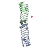
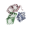
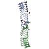
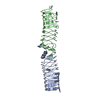
| Title | The Structure of MfpA (Rv3361c, C2 Crystal form). The Pentapeptide Repeat Protein from Mycobacterium tuberculosis Folds as A Right- handed Quadrilateral Beta-helix. | ||||||
 Components Components | PENTAPEPTIDE REPEAT FAMILY PROTEIN | ||||||
 Keywords Keywords | PENTAPEPTIDE REPEAT PROTEIN / PENTAPEPTIDE REPEAT PROTEINS / FLUROQUINOLONE RESISTANCE / DNA GYRASE / DNA MIMICRY / RIGHT-HANDED QUADRILATERAL BETA-HELIX | ||||||
| Function / homology |  Function and homology information Function and homology informationDNA topoisomerase type II (double strand cut, ATP-hydrolyzing) inhibitor activity / response to antibiotic / protein homodimerization activity Similarity search - Function | ||||||
| Biological species |   MYCOBACTERIUM TUBERCULOSIS (bacteria) MYCOBACTERIUM TUBERCULOSIS (bacteria) | ||||||
| Method |  X-RAY DIFFRACTION / MOLECULAR REPLACEMENT / Resolution: 2.2 Å X-RAY DIFFRACTION / MOLECULAR REPLACEMENT / Resolution: 2.2 Å | ||||||
 Authors Authors | Hegde, S.S. / Vetting, M.W. / Roderick, S.L. / Mitchenall, L.A. / Maxwell, A. / Takiff, H.E. / Blanchard, J.S. | ||||||
 Citation Citation | Journal: Science / Year: 2005 Title: A Fluroquinolone Resistance Protein from Mycobacterium Tuberculosis that Mimics DNA Authors: Hegde, S.S. / Vetting, M.W. / Roderick, S.L. / Mitchenall, L.A. / Maxwell, A. / Takiff, H.E. / Blanchard, J.S. | ||||||
| History |
|
- Structure visualization
| Structure viewer | Molecule: MolmilJmol/JSmol |
|---|
- Downloads & links
-Download
| PDBx/mmCIF format | 2bm4.cif.gz | 79.5 KB | Display | PDBx/mmCIF format |
|---|---|---|---|---|
| PDB format | pdb2bm4.ent.gz | 61 KB | Display | PDB format |
| PDBx/mmJSON format | 2bm4.json.gz | Tree view | PDBx/mmJSON format | |
| Others |  Other downloads Other downloads |
-Validation report
| Arichive directory | https://data.pdbj.org/pub/pdb/validation_reports/bm/2bm4ftp://data.pdbj.org/pub/pdb/validation_reports/bm/2bm4 | HTTPS FTP |
|---|
-Related structure data


-Links
 PDBj
PDBj- Assembly
| Deposited unit | 
| ||||||||
|---|---|---|---|---|---|---|---|---|---|
| 1 |
| ||||||||
| Unit cell |
|
-Components
| #1: Protein | Mass: 20325.859 Da / Num. of mol.: 2 Source method: isolated from a genetically manipulated source Source: (gene. exp.) MYCOBACTERIUM TUBERCULOSIS (bacteria) / Strain: H37RV / Plasmid: PET28A / Production host: #2: Water | ChemComp-HOH / |  Mass: 18.015 Da / Num. of mol.: 97 / Source method: isolated from a natural source / Formula: H2O Mass: 18.015 Da / Num. of mol.: 97 / Source method: isolated from a natural source / Formula: H2OSequence details | THREE RESIDUES FROM N-TERMINAL HIS-TAG REMAIN AFTER THROMBIN CLEAVAGE OF THE TAG. | |
|---|
-Experimental details
-Experiment
| Experiment | Method: X-RAY DIFFRACTION / Number of used crystals: 1 |
|---|
- Sample preparation
| Crystal | Density Matthews: 2.23 Å3/Da / Density % sol: 44.43 % | |||||||||||||||||||||||||||||||||||
|---|---|---|---|---|---|---|---|---|---|---|---|---|---|---|---|---|---|---|---|---|---|---|---|---|---|---|---|---|---|---|---|---|---|---|---|---|
| Crystal grow | Details: PROTEIN WAS CRYSTALLIZED FROM 20% PEG 400, 100 MM AMMONIUM CITRATE PH 5.5 | |||||||||||||||||||||||||||||||||||
| Crystal grow | *PLUS pH: 7.5 / Method: vapor diffusion | |||||||||||||||||||||||||||||||||||
| Components of the solutions | *PLUS
|
-Data collection
| Diffraction | Mean temperature: 93 K |
|---|---|
| Diffraction source | Source: ROTATING ANODE / Type: RIGAKU RU300 / Wavelength: 1.5418 |
| Detector | Type: MSC RAXIS IV / Detector: IMAGE PLATE |
| Radiation | Protocol: SINGLE WAVELENGTH / Monochromatic (M) / Laue (L): M / Scattering type: x-ray |
| Radiation wavelength | Wavelength: 1.5418 Å / Relative weight: 1 |
| Reflection | Resolution: 2.2→30 Å / Num. obs: 17270 / % possible obs: 93.3 % / Observed criterion σ(I): 0 / Redundancy: 2.6 % / Biso Wilson estimate: 23.2 Å2 / Rmerge(I) obs: 0.03 / Net I/σ(I): 19.8 |
| Reflection shell | Resolution: 2.2→2.28 Å / Redundancy: 2.2 % / Rmerge(I) obs: 0.13 / Mean I/σ(I) obs: 5.8 / % possible all: 85.5 |
| Reflection | *PLUS Rmerge(I) obs: 0.03 |
| Reflection shell | *PLUS % possible obs: 85.5 % / Rmerge(I) obs: 0.13 |
- Processing
| Software |
| ||||||||||||||||||||||||||||||||||||||||||||||||||||||||||||
|---|---|---|---|---|---|---|---|---|---|---|---|---|---|---|---|---|---|---|---|---|---|---|---|---|---|---|---|---|---|---|---|---|---|---|---|---|---|---|---|---|---|---|---|---|---|---|---|---|---|---|---|---|---|---|---|---|---|---|---|---|---|
| Refinement | Method to determine structure: MOLECULAR REPLACEMENT Starting model: PREVIOUS STRUCTURE DETERMINED BY SE-MET MAD Resolution: 2.2→30 Å / Cross valid method: THROUGHOUT / σ(F): 0 / Stereochemistry target values: MAXIMUM LIKELIHOOD Details: MISSING RESIDUES FROM THE N AND C- TERMINI OF THE SUBMITTED COORDINATES AS COMPARED TO THE SUBMITTED SEQUENCE ARE REGIONS WHICH EXHIBITED NO ELECTRON DENSITY AND THEREFORE THEREFORE WERE NOT MODELED.
| ||||||||||||||||||||||||||||||||||||||||||||||||||||||||||||
| Solvent computation | Solvent model: BULK SOLVENT / Bsol: 54.7426 Å2 / ksol: 0.362602 e/Å3 | ||||||||||||||||||||||||||||||||||||||||||||||||||||||||||||
| Displacement parameters | Biso mean: 26.6 Å2
| ||||||||||||||||||||||||||||||||||||||||||||||||||||||||||||
| Refinement step | Cycle: LAST / Resolution: 2.2→30 Å
| ||||||||||||||||||||||||||||||||||||||||||||||||||||||||||||
| Refine LS restraints |
| ||||||||||||||||||||||||||||||||||||||||||||||||||||||||||||
| LS refinement shell | Resolution: 2.2→2.3 Å / Rfactor Rfree: 0.301 / Rfactor Rwork: 0.203 / Total num. of bins used: 10 | ||||||||||||||||||||||||||||||||||||||||||||||||||||||||||||
| Software | *PLUS Name: CNS / Version: 1 / Classification: refinement | ||||||||||||||||||||||||||||||||||||||||||||||||||||||||||||
| Refinement | *PLUS Lowest resolution: 30 Å | ||||||||||||||||||||||||||||||||||||||||||||||||||||||||||||
| Solvent computation | *PLUS | ||||||||||||||||||||||||||||||||||||||||||||||||||||||||||||
| Displacement parameters | *PLUS |
