 Movie
Movie Controller


+ Open data

- Basic information
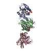
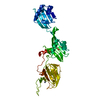
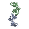
| Entry | Database: PDB / ID: 3wwk | ||||||
|---|---|---|---|---|---|---|---|
| Title | Crystal structure of CLEC-2 in complex with rhodocytin | ||||||
 Components Components |
| ||||||
 Keywords Keywords | SUGAR BINDING PROTEIN / C-type lectin fold / Carbohydrate binding / Podoplanin / Rhodocytin | ||||||
| Function / homology |  Function and homology information Function and homology informationregulation of cytokine activity / platelet formation / GPVI-mediated activation cascade / Heme signaling / defense response / transmembrane signaling receptor activity / toxin activity / carbohydrate binding / cell surface receptor signaling pathway / cell surface ...regulation of cytokine activity / platelet formation / GPVI-mediated activation cascade / Heme signaling / defense response / transmembrane signaling receptor activity / toxin activity / carbohydrate binding / cell surface receptor signaling pathway / cell surface / signal transduction / extracellular region / metal ion binding / plasma membrane Similarity search - Function | ||||||
| Biological species |  Homo sapiens (human) Homo sapiens (human) Calloselasma rhodostoma (Malayan pit viper) Calloselasma rhodostoma (Malayan pit viper) | ||||||
| Method |  X-RAY DIFFRACTION / SYNCHROTRON / MOLECULAR REPLACEMENT / Resolution: 2.98 Å X-RAY DIFFRACTION / SYNCHROTRON / MOLECULAR REPLACEMENT / Resolution: 2.98 Å | ||||||
 Authors Authors | Nagae, M. / Morita-Matsumoto, K. / Kato, M. / Kato-Kaneko, M. / Kato, Y. / Yamaguchi, Y. | ||||||
 Citation Citation | Journal: Structure / Year: 2014 Title: A Platform of C-type Lectin-like Receptor CLEC-2 for Binding O-Glycosylated Podoplanin and Nonglycosylated Rhodocytin Authors: Nagae, M. / Morita-Matsumoto, K. / Kato, M. / Kato-Kaneko, M. / Kato, Y. / Yamaguchi, Y. | ||||||
| History |
|
- Structure visualization
| Structure viewer | Molecule: MolmilJmol/JSmol |
|---|
- Downloads & links
-Download
| PDBx/mmCIF format | 3wwk.cif.gz | 307.6 KB | Display | PDBx/mmCIF format |
|---|---|---|---|---|
| PDB format | pdb3wwk.ent.gz | 251.4 KB | Display | PDB format |
| PDBx/mmJSON format | 3wwk.json.gz | Tree view | PDBx/mmJSON format | |
| Others |  Other downloads Other downloads |
-Validation report
| Arichive directory | https://data.pdbj.org/pub/pdb/validation_reports/ww/3wwkftp://data.pdbj.org/pub/pdb/validation_reports/ww/3wwk | HTTPS FTP |
|---|
-Related structure data
| Related structure data |  3wsrC 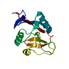 2c6uS  2vrpS C: citing same article ( S: Starting model for refinement |
|---|---|
| Similar structure data |


-Links
 PDBj
PDBj


- Assembly
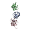
| Deposited unit | 
| ||||||||
|---|---|---|---|---|---|---|---|---|---|
| 1 | 
| ||||||||
| 2 | 
| ||||||||
| 3 | 
| ||||||||
| 4 | 
| ||||||||
| Unit cell |
|
-Components
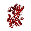
| #1: Protein | Mass: 15157.050 Da / Num. of mol.: 4 / Fragment: CLEC-2, UNP residues 96-221 / Mutation: C96S Source method: isolated from a genetically manipulated source Source: (gene. exp.) Homo sapiens (human) / Gene: CLEC1B, CLEC2, UNQ721/PRO1384 / Plasmid: pCold / Production host:  #2: Protein | Mass: 15812.376 Da / Num. of mol.: 4 / Source method: isolated from a natural source Source: (natural) Calloselasma rhodostoma (Malayan pit viper)References: UniProt: Q9I841 #3: Protein | Mass: 16790.094 Da / Num. of mol.: 4 / Source method: isolated from a natural source Source: (natural) Calloselasma rhodostoma (Malayan pit viper)References: UniProt: Q9I840 Has protein modification | Y | |
|---|
-Experimental details
-Experiment
| Experiment | Method: X-RAY DIFFRACTION / Number of used crystals: 1 |
|---|
- Sample preparation
| Crystal | Density Matthews: 2.75 Å3/Da / Density % sol: 55.24 % |
|---|---|
| Crystal grow | Temperature: 293 K / Method: vapor diffusion, sitting drop / pH: 7.6 Details: 0.1M Hepes (pH 7.6), 0.2M L-proline, 10% (w/v) PEG3350, VAPOR DIFFUSION, SITTING DROP, temperature 293K |
-Data collection
| Diffraction | Mean temperature: 95 K | |||||||||||||||
|---|---|---|---|---|---|---|---|---|---|---|---|---|---|---|---|---|
| Diffraction source | Source: SYNCHROTRON / Site: Photon Factory  / Beamline: BL-5A / Wavelength: 1 Å / Beamline: BL-5A / Wavelength: 1 Å | |||||||||||||||
| Detector | Type: ADSC QUANTUM 315r / Detector: CCD / Date: Jun 4, 2014 | |||||||||||||||
| Radiation | Monochromator: Numerical link type Si(111) double crystal monochromator Protocol: SINGLE WAVELENGTH / Monochromatic (M) / Laue (L): M / Scattering type: x-ray | |||||||||||||||
| Radiation wavelength | Wavelength: 1 Å / Relative weight: 1 | |||||||||||||||
| Reflection twin |
| |||||||||||||||
| Reflection | Resolution: 2.98→100 Å / Num. all: 41633 / Num. obs: 41508 / % possible obs: 99.7 % / Observed criterion σ(F): 0 / Redundancy: 3.6 % / Biso Wilson estimate: 44.3 Å2 / Rsym value: 0.099 / Net I/σ(I): 16.9 | |||||||||||||||
| Reflection shell | Resolution: 2.983→3.05 Å / Redundancy: 3.6 % / Mean I/σ(I) obs: 2.9 / Num. unique all: 2103 / Rsym value: 0.458 / % possible all: 99.9 |
- Processing
| Software |
| ||||||||||||||||||||||||||||||||||||||||||||||||||||||||||||||||||||||||||||||||||||||||||||||||||||||||||||||||||||||||||||||||||||||||||||||||||||||||||||||||||||||||||||||||||||||
|---|---|---|---|---|---|---|---|---|---|---|---|---|---|---|---|---|---|---|---|---|---|---|---|---|---|---|---|---|---|---|---|---|---|---|---|---|---|---|---|---|---|---|---|---|---|---|---|---|---|---|---|---|---|---|---|---|---|---|---|---|---|---|---|---|---|---|---|---|---|---|---|---|---|---|---|---|---|---|---|---|---|---|---|---|---|---|---|---|---|---|---|---|---|---|---|---|---|---|---|---|---|---|---|---|---|---|---|---|---|---|---|---|---|---|---|---|---|---|---|---|---|---|---|---|---|---|---|---|---|---|---|---|---|---|---|---|---|---|---|---|---|---|---|---|---|---|---|---|---|---|---|---|---|---|---|---|---|---|---|---|---|---|---|---|---|---|---|---|---|---|---|---|---|---|---|---|---|---|---|---|---|---|---|
| Refinement | Method to determine structure: MOLECULAR REPLACEMENT Starting model: 2C6U and 2VRP Resolution: 2.98→37.56 Å / Cor.coef. Fo:Fc: 0.853 / Cor.coef. Fo:Fc free: 0.79 / SU B: 32.825 / SU ML: 0.593 / Cross valid method: THROUGHOUT / ESU R Free: 0.118 / Stereochemistry target values: MAXIMUM LIKELIHOOD
| ||||||||||||||||||||||||||||||||||||||||||||||||||||||||||||||||||||||||||||||||||||||||||||||||||||||||||||||||||||||||||||||||||||||||||||||||||||||||||||||||||||||||||||||||||||||
| Solvent computation | Ion probe radii: 0.8 Å / Shrinkage radii: 0.8 Å / VDW probe radii: 1.2 Å / Solvent model: MASK | ||||||||||||||||||||||||||||||||||||||||||||||||||||||||||||||||||||||||||||||||||||||||||||||||||||||||||||||||||||||||||||||||||||||||||||||||||||||||||||||||||||||||||||||||||||||
| Displacement parameters | Biso mean: 60.923 Å2
| ||||||||||||||||||||||||||||||||||||||||||||||||||||||||||||||||||||||||||||||||||||||||||||||||||||||||||||||||||||||||||||||||||||||||||||||||||||||||||||||||||||||||||||||||||||||
| Refinement step | Cycle: LAST / Resolution: 2.98→37.56 Å
| ||||||||||||||||||||||||||||||||||||||||||||||||||||||||||||||||||||||||||||||||||||||||||||||||||||||||||||||||||||||||||||||||||||||||||||||||||||||||||||||||||||||||||||||||||||||
| Refine LS restraints |
|