 Movie
Movie Controller


+ Open data

- Basic information
| Entry | Database: PDB / ID: 6u2a | ||||||
|---|---|---|---|---|---|---|---|
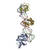
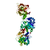
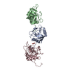
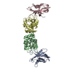
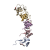
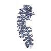
| Title | ShyA endopeptidase from Vibrio cholera (open form) | ||||||
 Components Components | ShyA endopeptidase | ||||||
 Keywords Keywords | HYDROLASE / M23 peptidase / peptidoglycan / crosslinks / periplasm | ||||||
| Function / homology |  Function and homology information Function and homology informationpeptidoglycan-based cell wall organization / peptidoglycan cross-bridge peptide endopeptidase activity / lysostaphin / unidimensional cell growth / peptidoglycan turnover / peptidoglycan binding / Hydrolases; Acting on peptide bonds (peptidases); Metalloendopeptidases / peptidoglycan catabolic process / peptidoglycan-based cell wall / metalloendopeptidase activity ...peptidoglycan-based cell wall organization / peptidoglycan cross-bridge peptide endopeptidase activity / lysostaphin / unidimensional cell growth / peptidoglycan turnover / peptidoglycan binding / Hydrolases; Acting on peptide bonds (peptidases); Metalloendopeptidases / peptidoglycan catabolic process / peptidoglycan-based cell wall / metalloendopeptidase activity / periplasmic space / hydrolase activity / proteolysis / zinc ion binding Similarity search - Function | ||||||
| Biological species |   Vibrio cholerae (bacteria) Vibrio cholerae (bacteria) | ||||||
| Method |  X-RAY DIFFRACTION / SYNCHROTRON / MOLECULAR REPLACEMENT / Resolution: 2.3 Å X-RAY DIFFRACTION / SYNCHROTRON / MOLECULAR REPLACEMENT / Resolution: 2.3 Å | ||||||
 Authors Authors | Saper, M.A. / Kelley, A. | ||||||
 Citation Citation | Journal: Proc.Natl.Acad.Sci.USA / Year: 2020 Title: Structural basis of peptidoglycan endopeptidase regulation. Authors: Shin, J.H. / Sulpizio, A.G. / Kelley, A. / Alvarez, L. / Murphy, S.G. / Fan, L. / Cava, F. / Mao, Y. / Saper, M.A. / Dorr, T. | ||||||
| History |
|
- Structure visualization
| Structure viewer | Molecule: MolmilJmol/JSmol |
|---|
- Downloads & links
-Download
| PDBx/mmCIF format | 6u2a.cif.gz | 180.6 KB | Display | PDBx/mmCIF format |
|---|---|---|---|---|
| PDB format | pdb6u2a.ent.gz | 116.8 KB | Display | PDB format |
| PDBx/mmJSON format | 6u2a.json.gz | Tree view | PDBx/mmJSON format | |
| Others |  Other downloads Other downloads |
-Validation report
| Arichive directory | https://data.pdbj.org/pub/pdb/validation_reports/u2/6u2aftp://data.pdbj.org/pub/pdb/validation_reports/u2/6u2a | HTTPS FTP |
|---|
-Related structure data
| Related structure data |  6ue4C  2guiS S: Starting model for refinement C: citing same article ( |
|---|---|
| Similar structure data |


-Links
 PDBj
PDBj- Assembly
| Deposited unit | 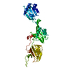
| ||||||||||
|---|---|---|---|---|---|---|---|---|---|---|---|
| 1 |
| ||||||||||
| Unit cell |
|
-Components
| #1: Protein | Mass: 45624.879 Da / Num. of mol.: 1 Source method: isolated from a genetically manipulated source Source: (gene. exp.) Vibrio cholerae (bacteria) / Gene: ShyA / Plasmid: pET28-VcShyA-His6Details (production host): Full-length ShyA followed by His6 Production host: References: UniProt: A0A0H6MKE7, UniProt: Q9KN86*PLUS, lysostaphin |
|---|---|
| #2: Chemical | ChemComp-ZN /   Mass: 65.409 Da / Num. of mol.: 1 / Source method: obtained synthetically / Formula: Zn Mass: 65.409 Da / Num. of mol.: 1 / Source method: obtained synthetically / Formula: Zn |
| #3: Water | ChemComp-HOH /  Mass: 18.015 Da / Num. of mol.: 10 / Source method: isolated from a natural source / Formula: H2O Mass: 18.015 Da / Num. of mol.: 10 / Source method: isolated from a natural source / Formula: H2O |
| Has ligand of interest | N |
-Experimental details
-Experiment
| Experiment | Method: X-RAY DIFFRACTION / Number of used crystals: 1 |
|---|
- Sample preparation
| Crystal | Density Matthews: 2.74 Å3/Da / Density % sol: 59.9 % / Description: elongated prisms |
|---|---|
| Crystal grow | Temperature: 295 K / Method: vapor diffusion, sitting drop / pH: 6 Details: Protein (0.5 microliter): 18 mg/ml in 20 mM Tris-HCl, 150 mM NaCl pH 7.6 Precipitant (0.5 microliter): 0.25 M sodium citrate, 10% polyethylene glycol 3350, 0.1 M Tris-HCl pH 6.0 PH range: 6-7.5 |
-Data collection
| Diffraction | Mean temperature: 125 K / Serial crystal experiment: N | |||||||||||||||||||||||||||
|---|---|---|---|---|---|---|---|---|---|---|---|---|---|---|---|---|---|---|---|---|---|---|---|---|---|---|---|---|
| Diffraction source | Source: SYNCHROTRON / Site: APS  / Beamline: 21-ID-D / Wavelength: 1.07811 Å / Beamline: 21-ID-D / Wavelength: 1.07811 Å | |||||||||||||||||||||||||||
| Detector | Type: DECTRIS EIGER X 9M / Detector: PIXEL / Date: Nov 18, 2018 | |||||||||||||||||||||||||||
| Radiation | Protocol: SINGLE WAVELENGTH / Monochromatic (M) / Laue (L): M / Scattering type: x-ray | |||||||||||||||||||||||||||
| Radiation wavelength | Wavelength: 1.07811 Å / Relative weight: 1 | |||||||||||||||||||||||||||
| Reflection | Resolution: 2.025→54.9 Å / Num. obs: 17391 / % possible obs: 54.1 % / Redundancy: 7 % / Biso Wilson estimate: 60.31 Å2 / CC1/2: 0.998 / Rmerge(I) obs: 0.055 / Rpim(I) all: 0.011 / Rrim(I) all: 0.059 / Net I/σ(I): 9.8 | |||||||||||||||||||||||||||
| Reflection shell | Diffraction-ID: 1
|
- Processing
| Software |
| |||||||||||||||||||||||||||||||||||||||||||||||||
|---|---|---|---|---|---|---|---|---|---|---|---|---|---|---|---|---|---|---|---|---|---|---|---|---|---|---|---|---|---|---|---|---|---|---|---|---|---|---|---|---|---|---|---|---|---|---|---|---|---|---|
| Refinement | Method to determine structure: MOLECULAR REPLACEMENT Starting model: 2GUI Resolution: 2.3→36.58 Å / SU ML: 0.2891 / Cross valid method: FREE R-VALUE / Phase error: 38.328
| |||||||||||||||||||||||||||||||||||||||||||||||||
| Solvent computation | Shrinkage radii: 0.9 Å / VDW probe radii: 1.11 Å | |||||||||||||||||||||||||||||||||||||||||||||||||
| Displacement parameters | Biso mean: 78.09 Å2 | |||||||||||||||||||||||||||||||||||||||||||||||||
| Refine analyze | Luzzati d res low obs: 40 Å | |||||||||||||||||||||||||||||||||||||||||||||||||
| Refinement step | Cycle: LAST / Resolution: 2.3→36.58 Å
| |||||||||||||||||||||||||||||||||||||||||||||||||
| Refine LS restraints |
| |||||||||||||||||||||||||||||||||||||||||||||||||
| LS refinement shell |
|
