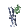
SHEET THE SHEET STRUCTURE OF THIS MOLECULE IS BIFURCATED. IN ORDER TO REPRESENT THIS FEATURE IN ... SHEET THE SHEET STRUCTURE OF THIS MOLECULE IS BIFURCATED. IN ORDER TO REPRESENT THIS FEATURE IN THE SHEET RECORDS BELOW, TWO SHEETS ARE DEFINED.
Mass: 18.015 Da / Num. of mol.: 44 / Source method: isolated from a natural source / Formula: H2O
Has protein modification
Y
Sequence details
FIRST THREE RESIDUES OF THE Q9I840 SEQUENCE OF CHAIN A ARE ABSENT FROM THE STRUCTURE. FIRST TWENTY- ...FIRST THREE RESIDUES OF THE Q9I840 SEQUENCE OF CHAIN A ARE ABSENT FROM THE STRUCTURE. FIRST TWENTY-THREE RESIDUES OF THE Q9I841 SEQUENCE OF CHAIN B ARE ABSENT FROM THE STRUCTURE, BECAUSE THESE RESIDUES ARE ABSENT FROM THE NATIVE PROTEIN (AS DETERMINED BY N-TERMINAL SEQUENCING, PMID 11728470)
-
Experimental details
-
Experiment
Experiment
Method: X-RAY DIFFRACTION / Number of used crystals: 1
-
Sample preparation
Crystal
Density Matthews: 2.77 Å3/Da / Density % sol: 55.65 % / Description: NONE
 Movie
Movie Controller


 Open data
 Basic information
 Components
 Keywords
 Function and homology information
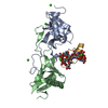
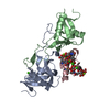
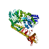
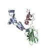
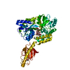
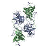
 CALLOSELASMA RHODOSTOMA (Malayan pit viper)
 X-RAY DIFFRACTION /
  Authors
 Citation
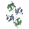
 Structure visualization
 Downloads & links
 Other downloads


 PDBj
PDBj


 Assembly
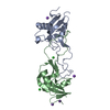

 Mass: 22.990 Da / Num. of mol.: 4 / Source method: obtained synthetically / Formula: Na
 Mass: 35.453 Da / Num. of mol.: 2 / Source method: obtained synthetically / Formula: Cl
 Mass: 18.015 Da / Num. of mol.: 44 / Source method: isolated from a natural source / Formula: H2O
 Sample preparation
 / Beamline: BM14 / Wavelength: 0.9762
  Processing