 Movie
Movie Controller

[English] 日本語
 Yorodumi
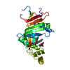
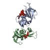
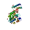
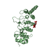
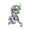
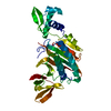
- PDB-1iod: CRYSTAL STRUCTURE OF THE COMPLEX BETWEEN THE COAGULATION FACTOR X... -
+ Open data

- Basic information
| Entry | Database: PDB / ID: 1iod | ||||||
|---|---|---|---|---|---|---|---|
| Title | CRYSTAL STRUCTURE OF THE COMPLEX BETWEEN THE COAGULATION FACTOR X BINDING PROTEIN FROM SNAKE VENOM AND THE GLA DOMAIN OF FACTOR X | ||||||
 Components Components |
| ||||||
 Keywords Keywords | HYDROLASE/HYDROLASE INHIBITOR / calcium bridging / domain swapping / HYDROLASE-HYDROLASE INHIBITOR COMPLEX | ||||||
| Function / homology |  Function and homology information Function and homology informationcoagulation factor Xa / blood coagulation / toxin activity / serine-type endopeptidase activity / calcium ion binding / proteolysis / : / extracellular region / metal ion binding Similarity search - Function | ||||||
| Biological species |  Deinagkistrodon acutus (Chinese moccasin) Deinagkistrodon acutus (Chinese moccasin) | ||||||
| Method |  X-RAY DIFFRACTION / SYNCHROTRON / MOLECULAR REPLACEMENT / Resolution: 2.3 Å X-RAY DIFFRACTION / SYNCHROTRON / MOLECULAR REPLACEMENT / Resolution: 2.3 Å | ||||||
 Authors Authors | Mizuno, H. / Fujimoto, Z. / Atoda, H. / Morita, T. | ||||||
 Citation Citation | Journal: Proc.Natl.Acad.Sci.Usa / Year: 2001 Title: Crystal structure of an anticoagulant protein in complex with the Gla domain of factor X. Authors: Mizuno, H. / Fujimoto, Z. / Atoda, H. / Morita, T. | ||||||
| History |
|
- Structure visualization
| Structure viewer | Molecule: MolmilJmol/JSmol |
|---|
- Downloads & links
-Download
| PDBx/mmCIF format | 1iod.cif.gz | 83.1 KB | Display | PDBx/mmCIF format |
|---|---|---|---|---|
| PDB format | pdb1iod.ent.gz | 62.3 KB | Display | PDB format |
| PDBx/mmJSON format | 1iod.json.gz | Tree view | PDBx/mmJSON format | |
| Others |  Other downloads Other downloads |
-Validation report
| Arichive directory | https://data.pdbj.org/pub/pdb/validation_reports/io/1iodftp://data.pdbj.org/pub/pdb/validation_reports/io/1iod | HTTPS FTP |
|---|
-Related structure data
| Related structure data | |
|---|---|
| Similar structure data |


-Links
 PDBj
PDBj


- Assembly
| Deposited unit | 
| |||||||||
|---|---|---|---|---|---|---|---|---|---|---|
| 1 |
| |||||||||
| 2 | 
| |||||||||
| 3 | 
| |||||||||
| Unit cell |
| |||||||||
| Components on special symmetry positions |
|
-Components
| #1: Protein | Mass: 14682.270 Da / Num. of mol.: 1 / Source method: isolated from a natural source / Source: (natural) Deinagkistrodon acutus (Chinese moccasin) / References: UniProt: Q9IAM1*PLUS | ||
|---|---|---|---|
| #2: Protein | Mass: 14580.163 Da / Num. of mol.: 1 / Source method: isolated from a natural source / Source: (natural) Deinagkistrodon acutus (Chinese moccasin) / References: UniProt: Q9DEF8*PLUS | ||
| #3: Protein/peptide | Mass: 5734.670 Da / Num. of mol.: 1 / Fragment: GLA DOMAIN(RESIDUES 41-84) / Source method: isolated from a natural source / Source: (natural) | ||
| #4: Chemical | ChemComp-CA /   Mass: 40.078 Da / Num. of mol.: 12 / Source method: obtained synthetically / Formula: Ca Mass: 40.078 Da / Num. of mol.: 12 / Source method: obtained synthetically / Formula: Ca#5: Water | ChemComp-HOH / |  Mass: 18.015 Da / Num. of mol.: 181 / Source method: isolated from a natural source / Formula: H2O Mass: 18.015 Da / Num. of mol.: 181 / Source method: isolated from a natural source / Formula: H2O |
-Experimental details
-Experiment
| Experiment | Method: X-RAY DIFFRACTION / Number of used crystals: 1 |
|---|
- Sample preparation
| Crystal | Density Matthews: 3.21 Å3/Da / Density % sol: 61.73 % | ||||||||||||||||||||||||
|---|---|---|---|---|---|---|---|---|---|---|---|---|---|---|---|---|---|---|---|---|---|---|---|---|---|
| Crystal grow | Temperature: 293 K / Method: vapor diffusion, hanging drop / pH: 7.9 Details: PEG8000, calcium chloride, pH 7.9, VAPOR DIFFUSION, HANGING DROP, temperature 293K | ||||||||||||||||||||||||
| Crystal grow | *PLUS Method: vapor diffusion | ||||||||||||||||||||||||
| Components of the solutions | *PLUS
|
-Data collection
| Diffraction | Mean temperature: 293 K |
|---|---|
| Diffraction source | Source: SYNCHROTRON / Site: Photon Factory  / Beamline: BL-6A / Wavelength: 1 Å / Beamline: BL-6A / Wavelength: 1 Å |
| Detector | Type: WEISSENBERG / Detector: DIFFRACTOMETER / Date: May 11, 1997 |
| Radiation | Protocol: SINGLE WAVELENGTH / Monochromatic (M) / Laue (L): M / Scattering type: x-ray |
| Radiation wavelength | Wavelength: 1 Å / Relative weight: 1 |
| Reflection | Resolution: 2.3→100 Å / Num. all: 109232 / Num. obs: 20411 / % possible obs: 96.4 % / Observed criterion σ(F): 2 / Observed criterion σ(I): 2 / Redundancy: 4.9 % / Biso Wilson estimate: 31.2 Å2 / Rmerge(I) obs: 0.081 / Net I/σ(I): 18.3 |
| Reflection shell | Resolution: 2.3→2.44 Å / Redundancy: 3.3 % / Rmerge(I) obs: 0.33 / Num. unique all: 3048 / % possible all: 90.3 |
| Reflection | *PLUS Lowest resolution: 100 Å / Num. measured all: 109232 |
| Reflection shell | *PLUS Highest resolution: 2.3 Å / % possible obs: 92.5 % / Num. unique obs: 3206 / Rmerge(I) obs: 0.33 |
- Processing
| Software |
| ||||||||||||||||||||
|---|---|---|---|---|---|---|---|---|---|---|---|---|---|---|---|---|---|---|---|---|---|
| Refinement | Method to determine structure: MOLECULAR REPLACEMENT Starting model: PDB ENTRY Resolution: 2.3→30 Å / Isotropic thermal model: Isotropic / Cross valid method: THROUGHOUT / σ(F): 2 / σ(I): 2 / Stereochemistry target values: Engh & Huber
| ||||||||||||||||||||
| Refine analyze |
| ||||||||||||||||||||
| Refinement step | Cycle: LAST / Resolution: 2.3→30 Å
| ||||||||||||||||||||
| Refine LS restraints |
| ||||||||||||||||||||
| LS refinement shell | Resolution: 2.3→2.44 Å / Rfactor Rfree error: 0.018
| ||||||||||||||||||||
| Refinement | *PLUS Highest resolution: 2.3 Å / Lowest resolution: 30 Å / σ(F): 2 | ||||||||||||||||||||
| Solvent computation | *PLUS | ||||||||||||||||||||
| Displacement parameters | *PLUS Biso mean: 24.3 Å2 | ||||||||||||||||||||
| Refine LS restraints | *PLUS Type: x_angle_deg / Dev ideal: 1.5 | ||||||||||||||||||||
| LS refinement shell | *PLUS Highest resolution: 2.3 Å / Rfactor Rfree: 0.301 / Rfactor Rwork: 0.302 |
