 Movie
Movie Controller

[English] 日本語
 Yorodumi
- PDB-1bj3: CRYSTAL STRUCTURE OF COAGULATION FACTOR IX-BINDING PROTEIN (IX-BP... -
+ Open data

- Basic information
| Entry | Database: PDB / ID: 1bj3 | ||||||
|---|---|---|---|---|---|---|---|
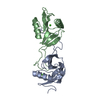
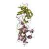
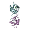
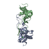
| Title | CRYSTAL STRUCTURE OF COAGULATION FACTOR IX-BINDING PROTEIN (IX-BP) FROM VENOM OF HABU SNAKE WITH A HETERODIMER OF C-TYPE LECTIN DOMAINS | ||||||
 Components Components |
| ||||||
 Keywords Keywords | COLLAGEN BINDING PROTEIN / COAGULATION FACTOR IX-BINDING / HETERODIMER / VENOM / HABU SNAKE / C-TYPE LECTIN SUPERFAMILY | ||||||
| Function / homology |  Function and homology information Function and homology information | ||||||
| Biological species |  Trimeresurus flavoviridis (habu) Trimeresurus flavoviridis (habu) | ||||||
| Method |  X-RAY DIFFRACTION / SYNCHROTRON / SIRAS / Resolution: 2.6 Å X-RAY DIFFRACTION / SYNCHROTRON / SIRAS / Resolution: 2.6 Å | ||||||
 Authors Authors | Mizuno, H. / Fujimoto, Z. / Koizumi, M. / Kano, H. / Atoda, H. / Morita, T. | ||||||
 Citation Citation | Journal: J.Mol.Biol. / Year: 1999 Title: Crystal structure of coagulation factor IX-binding protein from habu snake venom at 2.6 A: implication of central loop swapping based on deletion in the linker region. Authors: Mizuno, H. / Fujimoto, Z. / Koizumi, M. / Kano, H. / Atoda, H. / Morita, T. #1: Journal: Nat.Struct.Biol. / Year: 1997Title: Structure of Coagulation Factors Ix/X-Binding Protein, a Heterodimer of C-Type Lectin Domains Authors: Mizuno, H. / Fujimoto, Z. / Koizumi, M. / Kano, H. / Atoda, H. / Morita, T. #2: Journal: J.Biochem.(Tokyo) / Year: 1995Title: Blood Coagulation Factor Ix-Binding Protein from the Venom of Trimeresurus Flavoviridis: Purification and Characterization Authors: Atoda, H. / Ishikawa, M. / Yoshihara, E. / Sekiya, F. / Morita, T. | ||||||
| History |
|
- Structure visualization
| Structure viewer | Molecule: MolmilJmol/JSmol |
|---|
- Downloads & links
-Download
| PDBx/mmCIF format | 1bj3.cif.gz | 65.2 KB | Display | PDBx/mmCIF format |
|---|---|---|---|---|
| PDB format | pdb1bj3.ent.gz | 48.3 KB | Display | PDB format |
| PDBx/mmJSON format | 1bj3.json.gz | Tree view | PDBx/mmJSON format | |
| Others |  Other downloads Other downloads |
-Validation report
| Arichive directory | https://data.pdbj.org/pub/pdb/validation_reports/bj/1bj3ftp://data.pdbj.org/pub/pdb/validation_reports/bj/1bj3 | HTTPS FTP |
|---|
-Related structure data
| Similar structure data |
|---|


-Links
 PDBj
PDBj


- Assembly
| Deposited unit | 
| ||||||||
|---|---|---|---|---|---|---|---|---|---|
| 1 |
| ||||||||
| Unit cell |
|
-Components
| #1: Protein | Mass: 14655.184 Da / Num. of mol.: 1 / Fragment: C-TYPE LECTIN CRD DOMAIN / Source method: isolated from a natural source Details: DISULPHIDE DIMER BETWEEN A CYS79 AND B CYS75, PROTEIN COMPOSED OF HOMOLOGOUS SUBUNITS TO THE CARBOHYDRATE RECOGNITION DOMAIN OF C-TYPE LECTIN Source: (natural) Trimeresurus flavoviridis (habu) / Secretion: VENOM / References: PIR: JC4329, UniProt: Q7LZ71*PLUS | ||||||
|---|---|---|---|---|---|---|---|
| #2: Protein | Mass: 14455.071 Da / Num. of mol.: 1 / Fragment: C-TYPE LECTIN CRD DOMAIN / Source method: isolated from a natural source Details: DISULPHIDE DIMER BETWEEN A CYS79 AND B CYS75, PROTEIN COMPOSED OF HOMOLOGOUS SUBUNITS TO THE CARBOHYDRATE RECOGNITION DOMAIN OF C-TYPE LECTIN Source: (natural) Trimeresurus flavoviridis (habu) / Secretion: VENOM / References: UniProt: P23807 | ||||||
| #3: Chemical |   Mass: 40.078 Da / Num. of mol.: 2 / Source method: obtained synthetically / Formula: Ca Mass: 40.078 Da / Num. of mol.: 2 / Source method: obtained synthetically / Formula: Ca#4: Water | ChemComp-HOH / |  Mass: 18.015 Da / Num. of mol.: 83 / Source method: isolated from a natural source / Formula: H2O Mass: 18.015 Da / Num. of mol.: 83 / Source method: isolated from a natural source / Formula: H2OHas protein modification | Y | Sequence details | REFERENCE: MATURE PROTEIN LACKS INITIAL 23 RESIDUES, B CHAIN OF IX-BINDING PROTEIN IS THE SAME AS ...REFERENCE: MATURE PROTEIN LACKS INITIAL 23 RESIDUES, B CHAIN OF IX-BINDING PROTEIN IS THE SAME AS THAT OF FACTOR IX/FACTOR X-BINDING ANTICOAGUL | |
-Experimental details
-Experiment
| Experiment | Method: X-RAY DIFFRACTION / Number of used crystals: 2 |
|---|
- Sample preparation
| Crystal | Density Matthews: 2.3 Å3/Da / Density % sol: 47 % | ||||||||||||||||||||||||||||||||||||
|---|---|---|---|---|---|---|---|---|---|---|---|---|---|---|---|---|---|---|---|---|---|---|---|---|---|---|---|---|---|---|---|---|---|---|---|---|---|
| Crystal grow | pH: 7.8 Details: PROTEIN WAS CRYSTALLIZED BY MIXING WITH EQUAL VOLUME OF 60% AMMONIUM SULPHATE IN 20 MM TRI-HCL BUFFER AT PH 7.8 CONTAINING AND AT ROOM TEMPERATURE | ||||||||||||||||||||||||||||||||||||
| Crystal grow | *PLUS Temperature: 20 ℃ / Method: vapor diffusion, hanging drop | ||||||||||||||||||||||||||||||||||||
| Components of the solutions | *PLUS
|
-Data collection
| Diffraction | Mean temperature: 298 K |
|---|---|
| Diffraction source | Source: SYNCHROTRON / Site: Photon Factory  / Beamline: BL-6A / Wavelength: 1 / Beamline: BL-6A / Wavelength: 1 |
| Detector | Type: RIGAKU / Detector: IMAGE PLATE / Date: Jun 15, 1995 |
| Radiation | Protocol: SINGLE WAVELENGTH / Monochromatic (M) / Laue (L): M / Scattering type: x-ray |
| Radiation wavelength | Wavelength: 1 Å / Relative weight: 1 |
| Reflection | Resolution: 2.6→100 Å / Num. obs: 7253 / % possible obs: 90 % / Observed criterion σ(I): 3 / Redundancy: 4.1 % / Biso Wilson estimate: 25.1 Å2 / Rmerge(I) obs: 0.102 / Net I/σ(I): 12 |
| Reflection shell | Resolution: 2.6→2.72 Å / Redundancy: 2.3 % / Rmerge(I) obs: 0.145 / Mean I/σ(I) obs: 8.6 / % possible all: 74 |
| Reflection | *PLUS Num. measured all: 21947 |
- Processing
| Software |
| ||||||||||||||||||||||||||||||||||||||||||||||||||||||||||||||||||||||||||||||||
|---|---|---|---|---|---|---|---|---|---|---|---|---|---|---|---|---|---|---|---|---|---|---|---|---|---|---|---|---|---|---|---|---|---|---|---|---|---|---|---|---|---|---|---|---|---|---|---|---|---|---|---|---|---|---|---|---|---|---|---|---|---|---|---|---|---|---|---|---|---|---|---|---|---|---|---|---|---|---|---|---|---|
| Refinement | Method to determine structure: SIRAS / Resolution: 2.6→6 Å / Rfactor Rfree error: 0.014 / Data cutoff high absF: 10000000 / Data cutoff low absF: 0.001 / Isotropic thermal model: RESTRAINED / Cross valid method: THROUGHOUT / σ(F): 2
| ||||||||||||||||||||||||||||||||||||||||||||||||||||||||||||||||||||||||||||||||
| Displacement parameters | Biso mean: 17.3 Å2
| ||||||||||||||||||||||||||||||||||||||||||||||||||||||||||||||||||||||||||||||||
| Refine analyze |
| ||||||||||||||||||||||||||||||||||||||||||||||||||||||||||||||||||||||||||||||||
| Refinement step | Cycle: LAST / Resolution: 2.6→6 Å
| ||||||||||||||||||||||||||||||||||||||||||||||||||||||||||||||||||||||||||||||||
| Refine LS restraints |
| ||||||||||||||||||||||||||||||||||||||||||||||||||||||||||||||||||||||||||||||||
| LS refinement shell | Resolution: 2.6→2.75 Å / Rfactor Rfree error: 0.045 / Total num. of bins used: 6
| ||||||||||||||||||||||||||||||||||||||||||||||||||||||||||||||||||||||||||||||||
| Xplor file |
| ||||||||||||||||||||||||||||||||||||||||||||||||||||||||||||||||||||||||||||||||
| Software | *PLUS Name: X-PLOR / Version: 2.1 / Classification: refinement | ||||||||||||||||||||||||||||||||||||||||||||||||||||||||||||||||||||||||||||||||
| Refinement | *PLUS Highest resolution: 2.6 Å / % reflection Rfree: 5.6 % | ||||||||||||||||||||||||||||||||||||||||||||||||||||||||||||||||||||||||||||||||
| Solvent computation | *PLUS | ||||||||||||||||||||||||||||||||||||||||||||||||||||||||||||||||||||||||||||||||
| Displacement parameters | *PLUS Biso mean: 17.3 Å2 | ||||||||||||||||||||||||||||||||||||||||||||||||||||||||||||||||||||||||||||||||
| Refine LS restraints | *PLUS
| ||||||||||||||||||||||||||||||||||||||||||||||||||||||||||||||||||||||||||||||||
| LS refinement shell | *PLUS Rfactor Rfree: 0.33 / % reflection Rfree: 5.9 % / Rfactor Rwork: 0.234 |
