 Movie
Movie Controller

[English] 日本語
 Yorodumi
- PDB-1ukm: Crystal structure of EMS16, an Antagonist of collagen receptor in... -
+ Open data

- Basic information
| Entry | Database: PDB / ID: 1ukm | ||||||
|---|---|---|---|---|---|---|---|
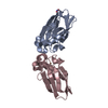
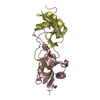
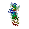
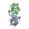
| Title | Crystal structure of EMS16, an Antagonist of collagen receptor integrin alpha2beta1 (GPIa/IIa) | ||||||
 Components Components |
| ||||||
 Keywords Keywords | TOXIN / Domain swapping / C-type lectin | ||||||
| Function / homology |  Function and homology information Function and homology information | ||||||
| Biological species |  Echis multisquamatus (snake) Echis multisquamatus (snake) | ||||||
| Method |  X-RAY DIFFRACTION / MOLECULAR REPLACEMENT / Resolution: 1.9 Å X-RAY DIFFRACTION / MOLECULAR REPLACEMENT / Resolution: 1.9 Å | ||||||
 Authors Authors | Horii, K. / Okuda, D. / Morita, T. / Mizuno, H. | ||||||
 Citation Citation | Journal: Biochemistry / Year: 2003 Title: Structural characterization of EMS16, an Antagonist of collagen receptor (GPIa/IIa) from the venom of Echis multisquamatus Authors: Horii, K. / Okuda, D. / Morita, T. / Mizuno, H. #1: Journal: J.BIOCHEM.(TOKYO) / Year: 2003Title: Characterization and Preliminary Crystallographic Studies of EMS16, an Antagonist of Collagen Receptor (GPIa/IIa) from the Venom of Echis multisquamatus Authors: Okuda, D. / Horii, K. / Mizuno, H. / Morita, T. #2: Journal: Biochemistry / Year: 2000Title: Isolation and characterization of EMS16, a C-lectin type protein from Echis multisquamatus venom, a potent and selective inhibitor of the alpha2beta1 integrin Authors: Marcinkiewicz, C. / Lobb, R.R. / Marcinkiewicz, M.M. / Daniel, J.L. / Smith, J.B. / Dangelmaier, C. / Weinreb, P.H. / Beacham, D.A. / Niewiarowski, S. | ||||||
| History |
|
- Structure visualization
| Structure viewer | Molecule: MolmilJmol/JSmol |
|---|
- Downloads & links
-Download
| PDBx/mmCIF format | 1ukm.cif.gz | 74.1 KB | Display | PDBx/mmCIF format |
|---|---|---|---|---|
| PDB format | pdb1ukm.ent.gz | 54.1 KB | Display | PDB format |
| PDBx/mmJSON format | 1ukm.json.gz | Tree view | PDBx/mmJSON format | |
| Others |  Other downloads Other downloads |
-Validation report
| Arichive directory | https://data.pdbj.org/pub/pdb/validation_reports/uk/1ukmftp://data.pdbj.org/pub/pdb/validation_reports/uk/1ukm | HTTPS FTP |
|---|
-Related structure data
| Related structure data |  1bj3S S: Starting model for refinement |
|---|---|
| Similar structure data |


-Links
 PDBj
PDBj


- Assembly
| Deposited unit | 
| ||||||||
|---|---|---|---|---|---|---|---|---|---|
| 1 |
| ||||||||
| Unit cell |
|
-Components
-Protein , 2 types, 2 molecules AB
| #1: Protein | Mass: 15889.537 Da / Num. of mol.: 1 / Fragment: RESIDUES 1-134 / Source method: isolated from a natural source / Source: (natural) Echis multisquamatus (snake) / Secretion: venom / References: UniProt: Q7T2Q1 |
|---|---|
| #2: Protein | Mass: 15121.384 Da / Num. of mol.: 1 / Fragment: RESIDUES 1-128 / Source method: isolated from a natural source / Source: (natural) Echis multisquamatus (snake) / Secretion: venom / References: UniProt: Q7T2Q0 |
-Sugars , 1 types, 1 molecules 
| #5: Sugar | ChemComp-NAG /  Type: D-saccharide, beta linking / Mass: 221.208 Da / Num. of mol.: 1 Type: D-saccharide, beta linking / Mass: 221.208 Da / Num. of mol.: 1Source method: isolated from a genetically manipulated source Formula: C8H15NO6 |
|---|
-Non-polymers , 3 types, 255 molecules 


| #3: Chemical | ChemComp-CL /  Mass: 35.453 Da / Num. of mol.: 1 / Source method: obtained synthetically / Formula: Cl Mass: 35.453 Da / Num. of mol.: 1 / Source method: obtained synthetically / Formula: Cl | ||
|---|---|---|---|
| #4: Chemical | ChemComp-GOL /  Mass: 92.094 Da / Num. of mol.: 4 / Source method: obtained synthetically / Formula: C3H8O3 Mass: 92.094 Da / Num. of mol.: 4 / Source method: obtained synthetically / Formula: C3H8O3#6: Water | ChemComp-HOH / | Mass: 18.015 Da / Num. of mol.: 250 / Source method: isolated from a natural source / Formula: H2O |
-Details
| Has protein modification | Y |
|---|---|
| Sequence details | The sequence database UNP Q7T2Q0 reports there is a conflict as Experimental Information. |
-Experimental details
-Experiment
| Experiment | Method: X-RAY DIFFRACTION / Number of used crystals: 1 |
|---|
- Sample preparation
| Crystal | Density Matthews: 2.39 Å3/Da / Density % sol: 48.1 % | ||||||||||||||||||||||||||||||||||||
|---|---|---|---|---|---|---|---|---|---|---|---|---|---|---|---|---|---|---|---|---|---|---|---|---|---|---|---|---|---|---|---|---|---|---|---|---|---|
| Crystal grow | Temperature: 293 K / Method: vapor diffusion, hanging drop / pH: 5.8 Details: PEG8000, potassium dihydrogen phosphate, glycerol, pH 5.8, VAPOR DIFFUSION, HANGING DROP, temperature 293K | ||||||||||||||||||||||||||||||||||||
| Crystal grow | *PLUS Temperature: 293 K / pH: 7.5 / Method: vapor diffusion, hanging drop | ||||||||||||||||||||||||||||||||||||
| Components of the solutions | *PLUS
|
-Data collection
| Diffraction | Mean temperature: 120 K |
|---|---|
| Diffraction source | Source: ROTATING ANODE / Type: RIGAKU / Wavelength: 1.5418 Å |
| Detector | Type: RIGAKU RAXIS IV / Detector: IMAGE PLATE / Date: Aug 12, 2002 / Details: Osmic confocal mirrors |
| Radiation | Monochromator: Osmic confocal / Protocol: SINGLE WAVELENGTH / Monochromatic (M) / Laue (L): M / Scattering type: x-ray |
| Radiation wavelength | Wavelength: 1.5418 Å / Relative weight: 1 |
| Reflection | Resolution: 1.9→29.71 Å / Num. obs: 26199 / % possible obs: 99.7 % / Observed criterion σ(I): 3 / Redundancy: 3.6 % / Biso Wilson estimate: 19.2 Å2 / Rmerge(I) obs: 0.031 / Net I/σ(I): 16.1 |
| Reflection shell | Resolution: 1.9→1.97 Å / Redundancy: 3.6 % / Rmerge(I) obs: 0.116 / Mean I/σ(I) obs: 5.6 / % possible all: 99.7 |
| Reflection | *PLUS Lowest resolution: 30 Å / Num. measured all: 91401 |
| Reflection shell | *PLUS % possible obs: 99.7 % |
- Processing
| Software |
| ||||||||||||||||||||||||||||||||||||||||||||||||||||||||||||||||||||||||||||||||
|---|---|---|---|---|---|---|---|---|---|---|---|---|---|---|---|---|---|---|---|---|---|---|---|---|---|---|---|---|---|---|---|---|---|---|---|---|---|---|---|---|---|---|---|---|---|---|---|---|---|---|---|---|---|---|---|---|---|---|---|---|---|---|---|---|---|---|---|---|---|---|---|---|---|---|---|---|---|---|---|---|---|
| Refinement | Method to determine structure: MOLECULAR REPLACEMENT Starting model: PDB ENTRY 1BJ3 Resolution: 1.9→18.36 Å / Rfactor Rfree error: 0.006 / Data cutoff high absF: 1271064.4 / Data cutoff low absF: 0 / Isotropic thermal model: RESTRAINED / Cross valid method: THROUGHOUT / σ(F): 0 / Stereochemistry target values: Engh & Huber
| ||||||||||||||||||||||||||||||||||||||||||||||||||||||||||||||||||||||||||||||||
| Solvent computation | Solvent model: FLAT MODEL / Bsol: 50.6729 Å2 / ksol: 0.394865 e/Å3 | ||||||||||||||||||||||||||||||||||||||||||||||||||||||||||||||||||||||||||||||||
| Displacement parameters | Biso mean: 27.7 Å2
| ||||||||||||||||||||||||||||||||||||||||||||||||||||||||||||||||||||||||||||||||
| Refine analyze |
| ||||||||||||||||||||||||||||||||||||||||||||||||||||||||||||||||||||||||||||||||
| Refinement step | Cycle: LAST / Resolution: 1.9→18.36 Å
| ||||||||||||||||||||||||||||||||||||||||||||||||||||||||||||||||||||||||||||||||
| Refine LS restraints |
| ||||||||||||||||||||||||||||||||||||||||||||||||||||||||||||||||||||||||||||||||
| LS refinement shell | Resolution: 1.9→2.02 Å / Rfactor Rfree error: 0.023 / Total num. of bins used: 6
| ||||||||||||||||||||||||||||||||||||||||||||||||||||||||||||||||||||||||||||||||
| Xplor file |
| ||||||||||||||||||||||||||||||||||||||||||||||||||||||||||||||||||||||||||||||||
| Refinement | *PLUS Lowest resolution: 20 Å / % reflection Rfree: 5 % / Rfactor Rfree: 0.234 | ||||||||||||||||||||||||||||||||||||||||||||||||||||||||||||||||||||||||||||||||
| Solvent computation | *PLUS | ||||||||||||||||||||||||||||||||||||||||||||||||||||||||||||||||||||||||||||||||
| Displacement parameters | *PLUS | ||||||||||||||||||||||||||||||||||||||||||||||||||||||||||||||||||||||||||||||||
| Refine LS restraints | *PLUS
|
