 Movie
Movie Controller


+ Open data

- Basic information
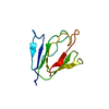
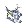
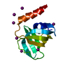
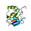
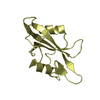
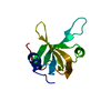
| Entry | Database: PDB / ID: 2i6v | ||||||
|---|---|---|---|---|---|---|---|
| Title | PDZ domain of EpsC from Vibrio cholerae, residues 219-305 | ||||||
 Components Components | General secretion pathway protein C | ||||||
 Keywords Keywords | PROTEIN TRANSPORT / MEMBRANE PROTEIN / EpsC / GspC / PDZ domain / Type 2 Secretion System / General Secretion Pathway | ||||||
| Function / homology |  Function and homology information Function and homology informationprotein secretion by the type II secretion system / type II protein secretion system complex / plasma membrane Similarity search - Function | ||||||
| Biological species |   Vibrio cholerae (bacteria) Vibrio cholerae (bacteria) | ||||||
| Method |  X-RAY DIFFRACTION / SAD / Resolution: 1.63 Å X-RAY DIFFRACTION / SAD / Resolution: 1.63 Å | ||||||
 Authors Authors | Korotkov, K.V. / Krumm, B. / Bagdasarian, M. / Hol, W.G.J. | ||||||
 Citation Citation | Journal: J.Mol.Biol. / Year: 2006 Title: Structural and Functional Studies of EpsC, a Crucial Component of the Type 2 Secretion System from Vibrio cholerae. Authors: Korotkov, K.V. / Krumm, B. / Bagdasarian, M. / Hol, W.G. | ||||||
| History |
|
- Structure visualization
| Structure viewer | Molecule: MolmilJmol/JSmol |
|---|
- Downloads & links
-Download
| PDBx/mmCIF format | 2i6v.cif.gz | 32 KB | Display | PDBx/mmCIF format |
|---|---|---|---|---|
| PDB format | pdb2i6v.ent.gz | 21.2 KB | Display | PDB format |
| PDBx/mmJSON format | 2i6v.json.gz | Tree view | PDBx/mmJSON format | |
| Others |  Other downloads Other downloads |
-Validation report
| Summary document | 2i6v_validation.pdf.gz | 406.5 KB | Display | wwPDB validaton report |
|---|---|---|---|---|
| Full document | 2i6v_full_validation.pdf.gz | 406.5 KB | Display | |
| Data in XML | 2i6v_validation.xml.gz | 6.8 KB | Display | |
| Data in CIF | 2i6v_validation.cif.gz | 8.9 KB | Display | |
| Arichive directory | https://data.pdbj.org/pub/pdb/validation_reports/i6/2i6vftp://data.pdbj.org/pub/pdb/validation_reports/i6/2i6v | HTTPS FTP |
-Related structure data


-Links
 PDBj
PDBj- Assembly
| Deposited unit | 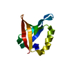
| ||||||||
|---|---|---|---|---|---|---|---|---|---|
| 1 |
| ||||||||
| Unit cell |
|
-Components
| #1: Protein | Mass: 9935.156 Da / Num. of mol.: 1 / Fragment: PDZ domain, residues 219-305 Source method: isolated from a genetically manipulated source Source: (gene. exp.) Vibrio cholerae (bacteria) / Gene: epsC / Plasmid: AVA0421 / Species (production host): Escherichia coli / Production host: |
|---|---|
| #2: Water | ChemComp-HOH /  Mass: 18.015 Da / Num. of mol.: 117 / Source method: isolated from a natural source / Formula: H2O Mass: 18.015 Da / Num. of mol.: 117 / Source method: isolated from a natural source / Formula: H2O |
-Experimental details
-Experiment
| Experiment | Method: X-RAY DIFFRACTION / Number of used crystals: 1 |
|---|
- Sample preparation
| Crystal | Density Matthews: 2.03 Å3/Da / Density % sol: 39.47 % |
|---|---|
| Crystal grow | Temperature: 294 K / Method: vapor diffusion / pH: 7.8 Details: 1.9M ammonium sulfate, 0.2M Li sulfate, 0.1M Tris pH 7.8, vapor diffusion, temperature 294K |
-Data collection
| Diffraction |
| |||||||||||||||||||||||||||||||||||||||||||||||||||||||||||||||||||||||||||||
|---|---|---|---|---|---|---|---|---|---|---|---|---|---|---|---|---|---|---|---|---|---|---|---|---|---|---|---|---|---|---|---|---|---|---|---|---|---|---|---|---|---|---|---|---|---|---|---|---|---|---|---|---|---|---|---|---|---|---|---|---|---|---|---|---|---|---|---|---|---|---|---|---|---|---|---|---|---|---|
| Diffraction source | Source: ROTATING ANODE / Type: OTHER / Wavelength: 1.5418 Å | |||||||||||||||||||||||||||||||||||||||||||||||||||||||||||||||||||||||||||||
| Detector | Type: MAC Science DIP-2030B / Detector: IMAGE PLATE / Date: May 3, 2005 / Details: Osmic optics | |||||||||||||||||||||||||||||||||||||||||||||||||||||||||||||||||||||||||||||
| Radiation |
| |||||||||||||||||||||||||||||||||||||||||||||||||||||||||||||||||||||||||||||
| Radiation wavelength | Wavelength: 1.5418 Å / Relative weight: 1 | |||||||||||||||||||||||||||||||||||||||||||||||||||||||||||||||||||||||||||||
| Reflection | Redundancy: 6.8 % / Av σ(I) over netI: 7.1 / Number: 74457 / Rmerge(I) obs: 0.07 / Rsym value: 0.07 / Χ2: 2.61 / D res high: 1.45 Å / D res low: 33.911 Å / Num. obs: 10912 / % possible obs: 72.8 | |||||||||||||||||||||||||||||||||||||||||||||||||||||||||||||||||||||||||||||
| Diffraction reflection shell | ID: 1
| |||||||||||||||||||||||||||||||||||||||||||||||||||||||||||||||||||||||||||||
| Reflection | Resolution: 1.63→50 Å / Num. obs: 10558 / % possible obs: 99.4 % / Redundancy: 5.4 % / Biso Wilson estimate: 17.5 Å2 / Rmerge(I) obs: 0.044 / Χ2: 2.614 / Net I/σ(I): 57.6 | |||||||||||||||||||||||||||||||||||||||||||||||||||||||||||||||||||||||||||||
| Reflection shell | Resolution: 1.63→1.69 Å / Redundancy: 5.1 % / Rmerge(I) obs: 0.187 / Mean I/σ(I) obs: 16.7 / Num. unique all: 1004 / Χ2: 3.484 / % possible all: 97.3 |
- Processing
| Software |
| ||||||||||||||||||||||||||||||||||||||||||||||||||||||||||||||||||||||||||||||||||||||||||
|---|---|---|---|---|---|---|---|---|---|---|---|---|---|---|---|---|---|---|---|---|---|---|---|---|---|---|---|---|---|---|---|---|---|---|---|---|---|---|---|---|---|---|---|---|---|---|---|---|---|---|---|---|---|---|---|---|---|---|---|---|---|---|---|---|---|---|---|---|---|---|---|---|---|---|---|---|---|---|---|---|---|---|---|---|---|---|---|---|---|---|---|
| Refinement | Method to determine structure: SAD / Resolution: 1.63→28.99 Å / Cor.coef. Fo:Fc: 0.963 / Cor.coef. Fo:Fc free: 0.952 / SU B: 1.732 / SU ML: 0.062 / Cross valid method: THROUGHOUT / σ(F): 0 / ESU R: 0.099 / ESU R Free: 0.097 / Stereochemistry target values: MAXIMUM LIKELIHOOD / Details: HYDROGENS HAVE BEEN ADDED IN THE RIDING POSITIONS
| ||||||||||||||||||||||||||||||||||||||||||||||||||||||||||||||||||||||||||||||||||||||||||
| Solvent computation | Ion probe radii: 0.8 Å / Shrinkage radii: 0.8 Å / VDW probe radii: 1.4 Å / Solvent model: MASK | ||||||||||||||||||||||||||||||||||||||||||||||||||||||||||||||||||||||||||||||||||||||||||
| Displacement parameters | Biso mean: 18.292 Å2
| ||||||||||||||||||||||||||||||||||||||||||||||||||||||||||||||||||||||||||||||||||||||||||
| Refinement step | Cycle: LAST / Resolution: 1.63→28.99 Å
| ||||||||||||||||||||||||||||||||||||||||||||||||||||||||||||||||||||||||||||||||||||||||||
| Refine LS restraints |
| ||||||||||||||||||||||||||||||||||||||||||||||||||||||||||||||||||||||||||||||||||||||||||
| LS refinement shell | Resolution: 1.63→1.672 Å / Total num. of bins used: 20
|
