 Movie
Movie Controller


+ Open data

- Basic information
| Entry | Database: PDB / ID: 2hhx | |||||||||
|---|---|---|---|---|---|---|---|---|---|---|
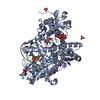
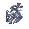
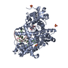
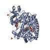
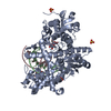
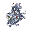
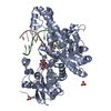
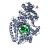
| Title | O6-methyl-guanine in the polymerase template preinsertion site | |||||||||
 Components Components |
| |||||||||
 Keywords Keywords | Transferase/DNA / DNA Polymerase I / DNA Replication / Klenow Fragment / Protein-DNA Complex / O6-Methyl-Guanine / Transferase-DNA COMPLEX | |||||||||
| Function / homology |  Function and homology information Function and homology information5'-3' exonuclease activity / 3'-5' exonuclease activity / DNA-templated DNA replication / double-strand break repair / DNA-directed DNA polymerase / DNA-directed DNA polymerase activity / nucleotide binding / DNA binding / metal ion binding Similarity search - Function | |||||||||
| Biological species |   Geobacillus stearothermophilus (bacteria) Geobacillus stearothermophilus (bacteria) | |||||||||
| Method |  X-RAY DIFFRACTION / FOURIER SYNTHESIS / Resolution: 2.26 Å X-RAY DIFFRACTION / FOURIER SYNTHESIS / Resolution: 2.26 Å | |||||||||
 Authors Authors | Warren, J.J. / Forsberg, L.J. / Beese, L.S. | |||||||||
 Citation Citation | Journal: Proc.Natl.Acad.Sci.Usa / Year: 2006 Title: The structural basis for the mutagenicity of O6-methyl-guanine lesions. Authors: Warren, J.J. / Forsberg, L.J. / Beese, L.S. | |||||||||
| History |
| |||||||||
| Remark 999 | SEQUENCE THE DNA POLYMERASE GENE WAS CLONED FROM AN ORGANISM THAT WAS CLASSIFIED AS A THERMOSTABLE ...SEQUENCE THE DNA POLYMERASE GENE WAS CLONED FROM AN ORGANISM THAT WAS CLASSIFIED AS A THERMOSTABLE STRAIN OF BACILLUS STEAROTHERMOPHILUS BY RIBOSOMAL RNA SEQUENCING. HOWEVER, THIS PARTICULAR GENE HAS MUCH GREATER HOMOLOGY WITH THE ANALOGOUS GENE FROM GEOBACILLUS KAUSTOPHILUS, UNP ACCESSION, Q5KWC1_GEOKA. |
- Structure visualization
| Structure viewer | Molecule: MolmilJmol/JSmol |
|---|
- Downloads & links
-Download
| PDBx/mmCIF format | 2hhx.cif.gz | 151 KB | Display | PDBx/mmCIF format |
|---|---|---|---|---|
| PDB format | pdb2hhx.ent.gz | 111.5 KB | Display | PDB format |
| PDBx/mmJSON format | 2hhx.json.gz | Tree view | PDBx/mmJSON format | |
| Others |  Other downloads Other downloads |
-Validation report
| Arichive directory | https://data.pdbj.org/pub/pdb/validation_reports/hh/2hhxftp://data.pdbj.org/pub/pdb/validation_reports/hh/2hhx | HTTPS FTP |
|---|
-Related structure data
| Related structure data |  2hhqC  2hhsC  2hhtC  2hhuC  2hhvC  2hhwC  2hvhC  2hviC  2hw3C  2hhy C: citing same article ( |
|---|---|
| Similar structure data |


-Links
 PDBj
PDBj


- Assembly
| Deposited unit | 
| ||||||||
|---|---|---|---|---|---|---|---|---|---|
| 1 |
| ||||||||
| Unit cell |
| ||||||||
| Details | The asymmetric unit contains one biological assembly. |
-Components
-DNA chain , 2 types, 2 molecules BC
| #1: DNA chain | Mass: 2691.774 Da / Num. of mol.: 1 / Source method: obtained synthetically / Details: Synthesized |
|---|---|
| #2: DNA chain | Mass: 4030.650 Da / Num. of mol.: 1 / Source method: obtained synthetically / Details: Synthesized |
-Protein / Sugars , 2 types, 3 molecules A
| #3: Protein | Mass: 66230.953 Da / Num. of mol.: 1 Fragment: residues 299-876 (Analogous to E Coli Klenow Fragment) Source method: isolated from a genetically manipulated source Source: (gene. exp.) Geobacillus stearothermophilus (bacteria)Gene: polA / Plasmid: PUC / Species (production host): Escherichia coli / Production host: References: UniProt: Q04957, UniProt: Q5KWC1*PLUS, DNA-directed DNA polymerase |
|---|---|
| #4: Polysaccharide |   Source method: isolated from a genetically manipulated source Details: oligosaccharide with reducing-end-to-reducing-end glycosidic bond References: sucrose |
-Non-polymers , 2 types, 316 molecules 

| #5: Chemical | ChemComp-SO4 /  Mass: 96.063 Da / Num. of mol.: 4 / Source method: obtained synthetically / Formula: SO4 Mass: 96.063 Da / Num. of mol.: 4 / Source method: obtained synthetically / Formula: SO4#6: Water | ChemComp-HOH / | Mass: 18.015 Da / Num. of mol.: 312 / Source method: isolated from a natural source / Formula: H2O |
|---|
-Experimental details
-Experiment
| Experiment | Method: X-RAY DIFFRACTION / Number of used crystals: 1 |
|---|
- Sample preparation
| Crystal | Density Matthews: 3.02 Å3/Da / Density % sol: 59.24 % | ||||||||||||||||||||||||||||||||
|---|---|---|---|---|---|---|---|---|---|---|---|---|---|---|---|---|---|---|---|---|---|---|---|---|---|---|---|---|---|---|---|---|---|
| Crystal grow | Temperature: 290 K / Method: vapor diffusion, hanging drop / pH: 5.8 Details: 45% saturated ammonium sulfate, 2.5% MPD, 100 mM MES, pH 5.8, VAPOR DIFFUSION, HANGING DROP, temperature 290K | ||||||||||||||||||||||||||||||||
| Components of the solutions |
|
-Data collection
| Diffraction | Mean temperature: 100 K |
|---|---|
| Diffraction source | Source: ROTATING ANODE / Type: RIGAKU RU200 / Wavelength: 1.5418 Å |
| Detector | Type: RIGAKU RAXIS II / Detector: IMAGE PLATE / Date: Oct 14, 2004 |
| Radiation | Monochromator: Yale Mirrors / Protocol: SINGLE WAVELENGTH / Monochromatic (M) / Laue (L): M / Scattering type: x-ray |
| Radiation wavelength | Wavelength: 1.5418 Å / Relative weight: 1 |
| Reflection | Resolution: 2.26→50 Å / Num. all: 39915 / Num. obs: 39915 / % possible obs: 95.8 % / Observed criterion σ(I): -3 / Redundancy: 2.8 % / Rsym value: 0.092 / Χ2: 1.041 / Net I/σ(I): 10.6 |
| Reflection shell | Resolution: 2.26→2.34 Å / Redundancy: 2.5 % / Mean I/σ(I) obs: 2.6 / Num. unique all: 3821 / Rsym value: 0.419 / Χ2: 1.05 / % possible all: 93.4 |
- Processing
| Software |
| |||||||||||||||||||||||||
|---|---|---|---|---|---|---|---|---|---|---|---|---|---|---|---|---|---|---|---|---|---|---|---|---|---|---|
| Refinement | Method to determine structure: FOURIER SYNTHESIS / Resolution: 2.26→50 Å / Isotropic thermal model: Isotropic / σ(F): 0
| |||||||||||||||||||||||||
| Solvent computation | Bsol: 35.423 Å2 | |||||||||||||||||||||||||
| Displacement parameters | Biso mean: 35.475 Å2
| |||||||||||||||||||||||||
| Refine analyze |
| |||||||||||||||||||||||||
| Refinement step | Cycle: LAST / Resolution: 2.26→50 Å
| |||||||||||||||||||||||||
| Refine LS restraints |
| |||||||||||||||||||||||||
| LS refinement shell | Resolution: 2.26→2.33 Å
| |||||||||||||||||||||||||
| Xplor file |
|
