 Movie
Movie Controller

[English] 日本語
 Yorodumi
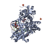
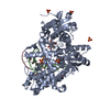
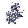
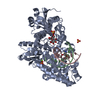
- PDB-3tap: Crystal Structure of Bacillus DNA Polymerase I Large Fragment Bou... -
+ Open data

- Basic information
| Entry | Database: PDB / ID: 3tap | |||||||||
|---|---|---|---|---|---|---|---|---|---|---|
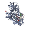
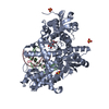
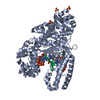
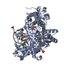
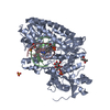
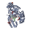
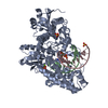
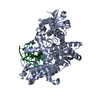
| Title | Crystal Structure of Bacillus DNA Polymerase I Large Fragment Bound to Duplex DNA with Cytosine-Adenine Mismatch at (n-3) Position | |||||||||
 Components Components |
| |||||||||
 Keywords Keywords | TRANSFERASE/DNA / DNA polymerase I / protein-DNA complex / TRANSFERASE-DNA complex | |||||||||
| Function / homology |  Function and homology information Function and homology informationTaq DNA Polymerase; Chain T, domain 4 / Taq DNA Polymerase; Chain T, domain 4 / Alpha-Beta Plaits - #370 / 5' to 3' exonuclease, C-terminal subdomain / Ribonuclease H-like superfamily/Ribonuclease H / DNA polymerase; domain 1 / Nucleotidyltransferase; domain 5 / Alpha-Beta Plaits / Up-down Bundle / 2-Layer Sandwich ...Taq DNA Polymerase; Chain T, domain 4 / Taq DNA Polymerase; Chain T, domain 4 / Alpha-Beta Plaits - #370 / 5' to 3' exonuclease, C-terminal subdomain / Ribonuclease H-like superfamily/Ribonuclease H / DNA polymerase; domain 1 / Nucleotidyltransferase; domain 5 / Alpha-Beta Plaits / Up-down Bundle / 2-Layer Sandwich / Orthogonal Bundle / Mainly Alpha / Alpha Beta Similarity search - Domain/homology | |||||||||
| Biological species |  Geobacillus (bacteria) Geobacillus (bacteria) | |||||||||
| Method |  X-RAY DIFFRACTION / SYNCHROTRON / FOURIER SYNTHESIS / Resolution: 1.655 Å X-RAY DIFFRACTION / SYNCHROTRON / FOURIER SYNTHESIS / Resolution: 1.655 Å | |||||||||
 Authors Authors | Wang, W. / Beese, L.S. | |||||||||
 Citation Citation | Journal: Proc.Natl.Acad.Sci.USA / Year: 2011 Title: Structural evidence for the rare tautomer hypothesis of spontaneous mutagenesis. Authors: Wang, W. / Hellinga, H.W. / Beese, L.S. #1: Journal: Cell(Cambridge,Mass.) / Year: 2004Title: Structures of mismatch replication errors observed in a DNA polymerase. Authors: Johnson, S.J. / Beese, L.S. | |||||||||
| History |
|
- Structure visualization
| Structure viewer | Molecule: MolmilJmol/JSmol |
|---|
- Downloads & links
-Download
| PDBx/mmCIF format | 3tap.cif.gz | 275.6 KB | Display | PDBx/mmCIF format |
|---|---|---|---|---|
| PDB format | pdb3tap.ent.gz | 215 KB | Display | PDB format |
| PDBx/mmJSON format | 3tap.json.gz | Tree view | PDBx/mmJSON format | |
| Others |  Other downloads Other downloads |
-Validation report
| Arichive directory | https://data.pdbj.org/pub/pdb/validation_reports/ta/3tapftp://data.pdbj.org/pub/pdb/validation_reports/ta/3tap | HTTPS FTP |
|---|
-Related structure data
| Related structure data |  3pv8C  3px0C  3px4C  3px6C  3tanC  3taqC  3tarC  3thvC  3ti0C  1l5uS C: citing same article ( S: Starting model for refinement |
|---|---|
| Similar structure data |


-Links
 PDBj
PDBj


- Assembly
| Deposited unit | 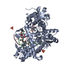
| ||||||||
|---|---|---|---|---|---|---|---|---|---|
| 1 |
| ||||||||
| Unit cell |
|
-Components
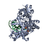
-DNA chain , 2 types, 2 molecules BC
| #2: DNA chain | Mass: 3632.382 Da / Num. of mol.: 1 / Source method: obtained synthetically / Details: Primer Strand |
|---|---|
| #3: DNA chain | Mass: 4923.204 Da / Num. of mol.: 1 / Source method: obtained synthetically / Details: Template Strand |
-Protein / Sugars , 2 types, 3 molecules A
| #1: Protein | Mass: 67518.297 Da / Num. of mol.: 1 / Fragment: Bacillus Fragment (UNP residues 285-876) Source method: isolated from a genetically manipulated source Source: (gene. exp.) Geobacillus (bacteria) / Plasmid: pET30a / Production host: |
|---|---|
| #4: Polysaccharide |   Source method: isolated from a genetically manipulated source Details: oligosaccharide with reducing-end-to-reducing-end glycosidic bond References: sucrose |
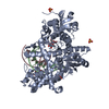
-Non-polymers , 3 types, 701 molecules 


| #5: Chemical | ChemComp-MG /  Mass: 24.305 Da / Num. of mol.: 1 / Source method: obtained synthetically / Formula: Mg Mass: 24.305 Da / Num. of mol.: 1 / Source method: obtained synthetically / Formula: Mg | ||
|---|---|---|---|
| #6: Chemical |  Mass: 96.063 Da / Num. of mol.: 3 / Source method: obtained synthetically / Formula: SO4 Mass: 96.063 Da / Num. of mol.: 3 / Source method: obtained synthetically / Formula: SO4#7: Water | ChemComp-HOH / | Mass: 18.015 Da / Num. of mol.: 697 / Source method: isolated from a natural source / Formula: H2O |
-Details
| Sequence details | THE AUTHORS STATE THAT THE GEOBACILLU |
|---|
-Experimental details
-Experiment
| Experiment | Method: X-RAY DIFFRACTION / Number of used crystals: 1 |
|---|
- Sample preparation
| Crystal | Density Matthews: 2.84 Å3/Da / Density % sol: 56.69 % |
|---|---|
| Crystal grow | Temperature: 290 K / Method: vapor diffusion, hanging drop / pH: 5.8 Details: 45-50% saturated ammonium sulfate, 2.5% MPD, 10 mM magnesium sulfate, 100 mM MES, pH 5.8, VAPOR DIFFUSION, HANGING DROP, temperature 290K |
-Data collection
| Diffraction | Mean temperature: 100 K | ||||||||||||||||||||||||||||||||||||||||||||||||||
|---|---|---|---|---|---|---|---|---|---|---|---|---|---|---|---|---|---|---|---|---|---|---|---|---|---|---|---|---|---|---|---|---|---|---|---|---|---|---|---|---|---|---|---|---|---|---|---|---|---|---|---|
| Diffraction source | Source: SYNCHROTRON / Site: APS  / Beamline: 22-BM / Wavelength: 0.97121 Å / Beamline: 22-BM / Wavelength: 0.97121 Å | ||||||||||||||||||||||||||||||||||||||||||||||||||
| Detector | Type: MARMOSAIC 225 mm CCD / Detector: CCD / Date: Oct 23, 2009 | ||||||||||||||||||||||||||||||||||||||||||||||||||
| Radiation | Monochromator: Rosenbaum-Rock double-crystal / Protocol: SINGLE WAVELENGTH / Monochromatic (M) / Laue (L): M / Scattering type: x-ray | ||||||||||||||||||||||||||||||||||||||||||||||||||
| Radiation wavelength | Wavelength: 0.97121 Å / Relative weight: 1 | ||||||||||||||||||||||||||||||||||||||||||||||||||
| Reflection | Resolution: 1.65→50 Å / Num. obs: 102589 / % possible obs: 98.1 % / Observed criterion σ(I): -3 / Rmerge(I) obs: 0.045 / Net I/σ(I): 19.92 | ||||||||||||||||||||||||||||||||||||||||||||||||||
| Reflection shell |
|
- Processing
| Software |
| |||||||||||||||||||||||||||||||||||||||||||||||||||||||||||||||||||||||||||||||||||||||||||||||||||||||||||||||||||||||||||||||||||||||||||||||||||||||||||||||||||||||||||||||||||||||||||||||||||||||||||||||||||||||||
|---|---|---|---|---|---|---|---|---|---|---|---|---|---|---|---|---|---|---|---|---|---|---|---|---|---|---|---|---|---|---|---|---|---|---|---|---|---|---|---|---|---|---|---|---|---|---|---|---|---|---|---|---|---|---|---|---|---|---|---|---|---|---|---|---|---|---|---|---|---|---|---|---|---|---|---|---|---|---|---|---|---|---|---|---|---|---|---|---|---|---|---|---|---|---|---|---|---|---|---|---|---|---|---|---|---|---|---|---|---|---|---|---|---|---|---|---|---|---|---|---|---|---|---|---|---|---|---|---|---|---|---|---|---|---|---|---|---|---|---|---|---|---|---|---|---|---|---|---|---|---|---|---|---|---|---|---|---|---|---|---|---|---|---|---|---|---|---|---|---|---|---|---|---|---|---|---|---|---|---|---|---|---|---|---|---|---|---|---|---|---|---|---|---|---|---|---|---|---|---|---|---|---|---|---|---|---|---|---|---|---|---|---|---|---|---|---|---|---|
| Refinement | Method to determine structure: FOURIER SYNTHESIS Starting model: PDB ENTRY 1L5U Resolution: 1.655→40.719 Å / Occupancy max: 1 / Occupancy min: 1 / SU ML: 0.32 / σ(F): 2.35 / Phase error: 19.76 / Stereochemistry target values: ML
| |||||||||||||||||||||||||||||||||||||||||||||||||||||||||||||||||||||||||||||||||||||||||||||||||||||||||||||||||||||||||||||||||||||||||||||||||||||||||||||||||||||||||||||||||||||||||||||||||||||||||||||||||||||||||
| Solvent computation | Shrinkage radii: 0.77 Å / VDW probe radii: 0.9 Å / Solvent model: FLAT BULK SOLVENT MODEL / Bsol: 47.157 Å2 / ksol: 0.446 e/Å3 | |||||||||||||||||||||||||||||||||||||||||||||||||||||||||||||||||||||||||||||||||||||||||||||||||||||||||||||||||||||||||||||||||||||||||||||||||||||||||||||||||||||||||||||||||||||||||||||||||||||||||||||||||||||||||
| Displacement parameters |
| |||||||||||||||||||||||||||||||||||||||||||||||||||||||||||||||||||||||||||||||||||||||||||||||||||||||||||||||||||||||||||||||||||||||||||||||||||||||||||||||||||||||||||||||||||||||||||||||||||||||||||||||||||||||||
| Refinement step | Cycle: LAST / Resolution: 1.655→40.719 Å
| |||||||||||||||||||||||||||||||||||||||||||||||||||||||||||||||||||||||||||||||||||||||||||||||||||||||||||||||||||||||||||||||||||||||||||||||||||||||||||||||||||||||||||||||||||||||||||||||||||||||||||||||||||||||||
| Refine LS restraints |
| |||||||||||||||||||||||||||||||||||||||||||||||||||||||||||||||||||||||||||||||||||||||||||||||||||||||||||||||||||||||||||||||||||||||||||||||||||||||||||||||||||||||||||||||||||||||||||||||||||||||||||||||||||||||||
| LS refinement shell |
|
