 Movie
Movie Controller

[English] 日本語
 Yorodumi
- PDB-3pv8: Crystal Structure of Bacillus DNA Polymerase I Large Fragment Bou... -
+ Open data

- Basic information
| Entry | Database: PDB / ID: 3pv8 | ||||||
|---|---|---|---|---|---|---|---|
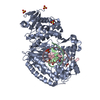
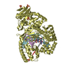
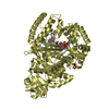
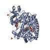
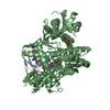
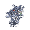
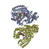
| Title | Crystal Structure of Bacillus DNA Polymerase I Large Fragment Bound to DNA and ddTTP-dA in Closed Conformation | ||||||
 Components Components |
| ||||||
 Keywords Keywords | TRANSFERASE/DNA / DNA Polymerase I / protein-DNA complex / Thymine-Adenine / closed conformation / TRANSFERASE-DNA complex | ||||||
| Function / homology |  Function and homology information Function and homology information5'-3' exonuclease activity / 3'-5' exonuclease activity / DNA-templated DNA replication / double-strand break repair / DNA-directed DNA polymerase / DNA-directed DNA polymerase activity / nucleotide binding / DNA binding / metal ion binding Similarity search - Function | ||||||
| Biological species |  Geobacillus kaustophilus (bacteria) Geobacillus kaustophilus (bacteria) | ||||||
| Method |  X-RAY DIFFRACTION / SYNCHROTRON / FOURIER SYNTHESIS / Resolution: 1.52 Å X-RAY DIFFRACTION / SYNCHROTRON / FOURIER SYNTHESIS / Resolution: 1.52 Å | ||||||
 Authors Authors | Wang, W. / Beese, L.S. | ||||||
 Citation Citation | Journal: Proc.Natl.Acad.Sci.USA / Year: 2011 Title: Structural evidence for the rare tautomer hypothesis of spontaneous mutagenesis. Authors: Wang, W. / Hellinga, H.W. / Beese, L.S. | ||||||
| History |
|
- Structure visualization
| Structure viewer | Molecule: MolmilJmol/JSmol |
|---|
- Downloads & links
-Download
| PDBx/mmCIF format | 3pv8.cif.gz | 530 KB | Display | PDBx/mmCIF format |
|---|---|---|---|---|
| PDB format | pdb3pv8.ent.gz | 428.6 KB | Display | PDB format |
| PDBx/mmJSON format | 3pv8.json.gz | Tree view | PDBx/mmJSON format | |
| Others |  Other downloads Other downloads |
-Validation report
| Arichive directory | https://data.pdbj.org/pub/pdb/validation_reports/pv/3pv8ftp://data.pdbj.org/pub/pdb/validation_reports/pv/3pv8 | HTTPS FTP |
|---|
-Related structure data
| Related structure data |  3px0C  3px4C  3px6C  3tanC  3tapC  3taqC  3tarC 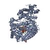 3thvC 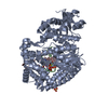 3ti0C  2hviS C: citing same article ( S: Starting model for refinement |
|---|---|
| Similar structure data |


-Links
 PDBj
PDBj


- Assembly
| Deposited unit | 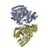
| ||||||||
|---|---|---|---|---|---|---|---|---|---|
| 1 | 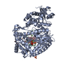
| ||||||||
| 2 | 
| ||||||||
| Unit cell |
|
-Components
-Protein , 1 types, 2 molecules AD
| #1: Protein | Mass: 67490.289 Da / Num. of mol.: 2 Fragment: Bacillus Fragment (analogous to E. coli Klenow Fragment) Mutation: D598A, F710Y Source method: isolated from a genetically manipulated source Source: (gene. exp.) Geobacillus kaustophilus (bacteria) / Gene: polA, GK2730 / Plasmid: pET30a / Production host: |
|---|
-DNA chain , 2 types, 4 molecules BECF
| #2: DNA chain | Mass: 2650.762 Da / Num. of mol.: 2 / Fragment: DNA primer strand / Source method: obtained synthetically #3: DNA chain | Mass: 4024.649 Da / Num. of mol.: 2 / Fragment: DNA template strand / Source method: obtained synthetically |
|---|
-Non-polymers , 4 types, 1830 molecules 


| #4: Chemical |  Mass: 24.305 Da / Num. of mol.: 2 / Source method: obtained synthetically / Formula: Mg Mass: 24.305 Da / Num. of mol.: 2 / Source method: obtained synthetically / Formula: Mg#5: Chemical |  Type: DNA OH 3 prime terminus / Mass: 466.169 Da / Num. of mol.: 2 / Source method: obtained synthetically / Formula: C10H17N2O13P3 Type: DNA OH 3 prime terminus / Mass: 466.169 Da / Num. of mol.: 2 / Source method: obtained synthetically / Formula: C10H17N2O13P3#6: Chemical |  Mass: 96.063 Da / Num. of mol.: 3 / Source method: obtained synthetically / Formula: SO4 Mass: 96.063 Da / Num. of mol.: 3 / Source method: obtained synthetically / Formula: SO4#7: Water | ChemComp-HOH / | Mass: 18.015 Da / Num. of mol.: 1823 / Source method: isolated from a natural source / Formula: H2O |
|---|
-Details
| Sequence details | AUTHORS STATE THAT THE PROTEIN WAS ISOLATED FROM A STRAIN OF GEOBACILLUS SPECIES WHOSE SEQUENCE IS ...AUTHORS STATE THAT THE PROTEIN WAS ISOLATED FROM A STRAIN OF GEOBACILLU |
|---|
-Experimental details
-Experiment
| Experiment | Method: X-RAY DIFFRACTION / Number of used crystals: 1 |
|---|
- Sample preparation
| Crystal | Density Matthews: 2.6 Å3/Da / Density % sol: 52.72 % | ||||||||||||||||||||||||||||||||||||
|---|---|---|---|---|---|---|---|---|---|---|---|---|---|---|---|---|---|---|---|---|---|---|---|---|---|---|---|---|---|---|---|---|---|---|---|---|---|
| Crystal grow | Temperature: 290 K / Method: vapor diffusion, hanging drop / pH: 5.8 Details: Ammonium Sulfate, MPD, MgSO4, MES, pH 5.8, vapor diffusion, hanging drop, temperature 290K | ||||||||||||||||||||||||||||||||||||
| Components of the solutions |
|
-Data collection
| Diffraction source | Source: SYNCHROTRON / Site: APS  / Beamline: 22-BM / Wavelength: 0.97121 Å / Beamline: 22-BM / Wavelength: 0.97121 Å | ||||||||||||||||||||||||||||||||||||||||||||||||||
|---|---|---|---|---|---|---|---|---|---|---|---|---|---|---|---|---|---|---|---|---|---|---|---|---|---|---|---|---|---|---|---|---|---|---|---|---|---|---|---|---|---|---|---|---|---|---|---|---|---|---|---|
| Detector | Type: MARMOSAIC 225 mm CCD / Detector: CCD / Date: Oct 23, 2009 | ||||||||||||||||||||||||||||||||||||||||||||||||||
| Radiation | Protocol: SINGLE WAVELENGTH / Monochromatic (M) / Laue (L): M / Scattering type: x-ray | ||||||||||||||||||||||||||||||||||||||||||||||||||
| Radiation wavelength | Wavelength: 0.97121 Å / Relative weight: 1 | ||||||||||||||||||||||||||||||||||||||||||||||||||
| Reflection | Resolution: 1.52→100 Å / Num. obs: 231085 / % possible obs: 97.5 % / Observed criterion σ(I): -3 / Rmerge(I) obs: 0.069 / Net I/σ(I): 14.94 | ||||||||||||||||||||||||||||||||||||||||||||||||||
| Reflection shell |
|
- Processing
| Software |
| |||||||||||||||||||||||||||||||||||||||||||||||||||||||||||||||||||||||||||||||||||||||||||||||||||||||||||||||||||||||||||||||||||||||||||||||||||||||||||||||||||||||||||||||||||||||||||||||||||||||||||||||||||||||||
|---|---|---|---|---|---|---|---|---|---|---|---|---|---|---|---|---|---|---|---|---|---|---|---|---|---|---|---|---|---|---|---|---|---|---|---|---|---|---|---|---|---|---|---|---|---|---|---|---|---|---|---|---|---|---|---|---|---|---|---|---|---|---|---|---|---|---|---|---|---|---|---|---|---|---|---|---|---|---|---|---|---|---|---|---|---|---|---|---|---|---|---|---|---|---|---|---|---|---|---|---|---|---|---|---|---|---|---|---|---|---|---|---|---|---|---|---|---|---|---|---|---|---|---|---|---|---|---|---|---|---|---|---|---|---|---|---|---|---|---|---|---|---|---|---|---|---|---|---|---|---|---|---|---|---|---|---|---|---|---|---|---|---|---|---|---|---|---|---|---|---|---|---|---|---|---|---|---|---|---|---|---|---|---|---|---|---|---|---|---|---|---|---|---|---|---|---|---|---|---|---|---|---|---|---|---|---|---|---|---|---|---|---|---|---|---|---|---|---|
| Refinement | Method to determine structure: FOURIER SYNTHESIS Starting model: PDB entry 2HVI with insertion site base pair, metal ions, and protein residues 681-727 deleted. Resolution: 1.52→88.422 Å / Cor.coef. Fo:Fc: 0.95 / Cor.coef. Fo:Fc free: 0.936 / Occupancy max: 1 / Occupancy min: 0.5 / SU ML: 0.36 / σ(F): 2.35 / Phase error: 19.45 / Stereochemistry target values: ML
| |||||||||||||||||||||||||||||||||||||||||||||||||||||||||||||||||||||||||||||||||||||||||||||||||||||||||||||||||||||||||||||||||||||||||||||||||||||||||||||||||||||||||||||||||||||||||||||||||||||||||||||||||||||||||
| Solvent computation | Shrinkage radii: 0.77 Å / VDW probe radii: 0.9 Å / Solvent model: FLAT BULK SOLVENT MODEL / Bsol: 44.623 Å2 / ksol: 0.411 e/Å3 | |||||||||||||||||||||||||||||||||||||||||||||||||||||||||||||||||||||||||||||||||||||||||||||||||||||||||||||||||||||||||||||||||||||||||||||||||||||||||||||||||||||||||||||||||||||||||||||||||||||||||||||||||||||||||
| Displacement parameters |
| |||||||||||||||||||||||||||||||||||||||||||||||||||||||||||||||||||||||||||||||||||||||||||||||||||||||||||||||||||||||||||||||||||||||||||||||||||||||||||||||||||||||||||||||||||||||||||||||||||||||||||||||||||||||||
| Refinement step | Cycle: LAST / Resolution: 1.52→88.422 Å
| |||||||||||||||||||||||||||||||||||||||||||||||||||||||||||||||||||||||||||||||||||||||||||||||||||||||||||||||||||||||||||||||||||||||||||||||||||||||||||||||||||||||||||||||||||||||||||||||||||||||||||||||||||||||||
| Refine LS restraints |
| |||||||||||||||||||||||||||||||||||||||||||||||||||||||||||||||||||||||||||||||||||||||||||||||||||||||||||||||||||||||||||||||||||||||||||||||||||||||||||||||||||||||||||||||||||||||||||||||||||||||||||||||||||||||||
| LS refinement shell |
|
